
🇦🇪 DHA · DOH · MOHAP
Specialist Oral & maxillofacial surgery United Arab Emirates
Specialist dentists preparing for the Oral & maxillofacial surgery exams of DHA, DOH, Qatar DHP and SCFHS
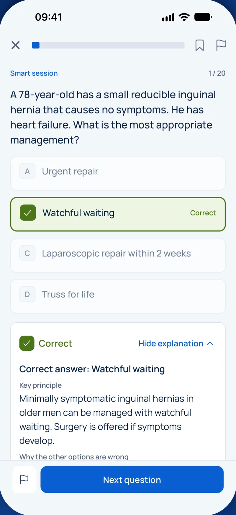
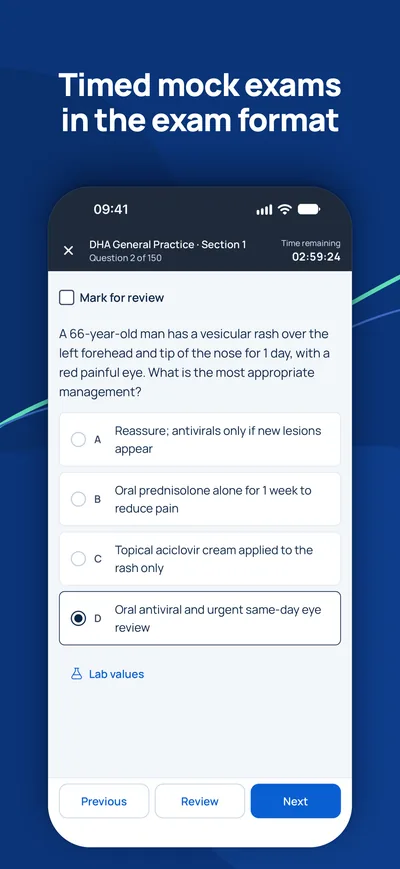
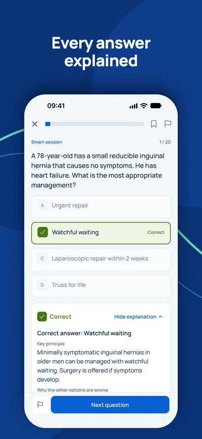
سؤال نموذجي
Which radiographic sign on a panoramic radiograph is most strongly associated with direct contact between a mandibular third molar root and the inferior alveolar canal?
Keys: 1–5 or A–E to choose, Enter to check and continue, F to mark.
Original MediFlow content, not official exam questions. Medical review in progress.
Syllabus and high-yield notes
1. Exodontia & impactions · 15 % الوزن في الامتحان · 5 lessons+
- NICE TA1 restricts prophylactic removal of pathology-free third molars; indications include unrestorable caries, untreatable pulpal or periapical pathology, cellulitis, abscess, osteomyelitis, internal/external resorption, fracture, cyst or tumour, and a second episode (or one severe episode) of pericoronitis.
- Rood's radiographic signs of close root–canal relationship: darkening of the root, deflection of the root, narrowing of the root, dark and bifid apex; and for the canal: interruption of the white line, diversion and narrowing of the canal. Diversion, darkening of the root and interruption of the white line carry the most risk.
- CBCT is indicated when a panoramic image shows a high-risk sign, to plan surgery or coronectomy; it is not a routine pre-extraction investigation.
- Winter's classification describes angulation (mesioangular, vertical, distoangular, horizontal); mesioangular is commonest and easiest in the mandible, distoangular hardest. In the maxilla the distoangular tooth is the easiest.
- Most lingual nerve injuries after third molar surgery recover within months; persistent complete anaesthesia beyond about 3 months with no recovery is the usual trigger for referral for microsurgical exploration.
- An oroantral communication under about 2 mm usually closes with a clot and sinus precautions; larger defects need primary closure (buccal advancement flap, buccal fat pad or palatal rotation flap).
- A fractured maxillary tuberosity that remains attached to periosteum is reduced and splinted, and the tooth is removed surgically 6–8 weeks later.
- Coronectomy leaves vital roots at least 3 mm below the crest; root migration is expected and re-operation is needed in a small minority. It is contraindicated with mobile or infected roots and active pericoronal pathology involving the root.
- Principles of exodontia: assessment, instruments and technique – Exodontia is the removal of teeth. At specialist level the examiner is not interested in whether you can use forceps; they want to know whether you can predict difficulty, choose the right technique before you start, and recognise when a simple extraction should become a surgical one. Most complications are decided in the planning phase, not in the chair.
- Impacted third molars: indications, classification, imaging and surgery – An impacted tooth is one that cannot erupt into a functional position because of bone, soft tissue or another tooth in its path. Lower third molars are the most commonly impacted teeth. The key specialist skills are deciding whether the tooth should come out at all, describing it in standard terms, judging the risk to the inferior alveolar nerve, and choosing the operation that minimises harm.
- Complications of exodontia: nerves, sinus, displaced teeth and dry socket – Complications are the core of specialist exams because they test judgement. For each one you should know how to prevent it, how to recognise it, what to do on the day, and when to refer.
- Local anaesthesia and pain management in oral surgery – Almost every oral surgical procedure starts with local anaesthesia, and many problems that candidates are asked about (failed blocks, toxicity, nerve injury, postoperative pain) come back to how well the anaesthetic and analgesic plan was built. This chapter explains how local anaesthetics work, how to choose the agent and dose, how the main blocks are given, why they fail, and how to control pain after surgery.
- Preprosthetic, implant and bone augmentation surgery – After teeth are lost, the alveolar ridge resorbs. The greatest loss happens in the first six to twelve months, and horizontal width is lost faster than height. Preprosthetic surgery prepares the mouth for a denture or implants: it removes obstacles, deepens the sulcus and, today mostly, rebuilds bone so that implants can be placed in the right prosthetic position. This chapter covers soft and hard tissue preprosthetic procedures, socket preservation, the basics of implant surgery, and the main augmentation techniques with their complications.
2. Infections · 15 % الوزن في الامتحان · 5 lessons+
- Odontogenic infections are polymicrobial (viridans streptococci, Prevotella, Fusobacterium, Peptostreptococcus). Source control by drainage and removal of the cause is the primary treatment; antibiotics are adjuncts.
- Flynn severity scores rank spaces: low risk (vestibular, buccal, canine, infraorbital), moderate (submandibular, submental, sublingual, pterygomandibular, submasseteric, temporal), severe (lateral pharyngeal, retropharyngeal, pretracheal, mediastinum, danger space).
- Ludwig angina is bilateral cellulitis of the submandibular, sublingual and submental spaces; airway protection (awake fibreoptic intubation or tracheostomy under local) comes before incision and drainage.
- Amoxicillin (or phenoxymethylpenicillin) is first line; add metronidazole for spreading infection; clindamycin is used in penicillin allergy but carries C. difficile risk and rising resistance.
- Cavernous sinus thrombosis from upper anterior teeth spreads via valveless facial, angular and ophthalmic veins or via the pterygoid plexus; signs are proptosis, chemosis, ophthalmoplegia (CN VI first) and spread to the other eye.
- Descending necrotising mediastinitis is diagnosed on contrast CT of neck and chest; it needs urgent cervical and thoracic drainage and broad-spectrum antibiotics, with mortality still substantial.
- Chronic suppurative osteomyelitis of the mandible needs sequestrectomy, decortication where indicated and prolonged culture-guided antibiotics; Garré osteomyelitis (proliferative periostitis) in children shows onion-skin periosteal bone and resolves after the dental cause is treated.
- Necrotising fasciitis of the neck shows crepitus, dusky skin, pain out of proportion and systemic toxicity; treatment is emergency radical debridement plus broad-spectrum antibiotics.
- Odontogenic infection: microbiology, stages and fascial spaces – Odontogenic infections start from a tooth or its supporting tissues, most often a necrotic pulp, a periodontal pocket or pericoronitis. Pus follows the path of least resistance. It perforates the thinnest cortical plate near the root apex and then enters whichever soft-tissue compartment lies on that side of the nearest muscle attachment. If you know where the apex sits relative to muscle attachments, you can predict which space is involved.
- Managing odontogenic infection: airway, drainage and antibiotics – Management follows a fixed order. First judge severity and the airway. Then decide where the patient is treated (chair or hospital). Then achieve source control by draining pus and removing the cause. Antibiotics support this, they do not replace it. Finally, review: if the patient is not improving after 48 to 72 hours, look for the reason rather than simply switching drugs.
- Life-threatening spread, osteomyelitis and chronic infections – Most odontogenic infections settle with drainage and removal of the cause. A small number spread into the neck, chest, veins or skull, or become chronic in bone. These are rare in practice but common in exams because they test recognition of dangerous patterns.
- Maxillary sinus, salivary gland and specific infections – Not every infection an oral surgeon sees starts in a tooth abscess and spreads into a fascial space. Three other groups matter in practice and in exams: infections of the maxillary sinus (including those caused by teeth and by surgery), infections of the salivary glands, and specific infections with their own organisms and patterns, such as actinomycosis, tuberculosis, fungal and viral infections. This chapter explains how to recognise each one and what makes its management different.
- Antibiotic prophylaxis and stewardship in oral surgery – Antibiotics save lives in spreading infection, but most dental pain does not need them, and inappropriate prescribing drives resistance, Clostridioides difficile infection and allergic reactions. Dentists write a meaningful share of all outpatient antibiotic prescriptions. Exams test two linked ideas: when antibiotics are actually indicated (treatment and prophylaxis), and how to choose drug, dose and duration responsibly. This chapter brings both together.
3. Trauma · 15 % الوزن في الامتحان · 5 lessons+
- Airway, cervical spine and haemorrhage (ATLS primary survey) come before any facial fracture management; bilateral parasymphyseal fractures can let the tongue fall back.
- Champy's ideal lines of osteosynthesis: one miniplate below the apices in the angle along the external oblique ridge, and two plates at the symphysis/parasymphysis (tension and compression zones).
- Load-bearing osteosynthesis (reconstruction plates) is needed for comminuted, atrophic edentulous or defect fractures; load-sharing miniplates suit simple fractures with good bone contact.
- Retrobulbar haemorrhage is an emergency: pain, proptosis, tense globe, reduced acuity and an afferent pupil defect; do a lateral canthotomy and cantholysis immediately.
- A white-eyed blowout fracture in a child (trapdoor with muscle entrapment, oculocardiac reflex with bradycardia, nausea) needs urgent release within 24–48 hours.
- Condylar fractures in children are usually treated closed with early mobilisation because of remodelling potential; ORIF is considered in adults with displaced subcondylar fractures, loss of ramus height or malocclusion.
- IADT 2020: avulsed permanent teeth with closed apices replanted within 60 minutes dry time receive a flexible splint for 2 weeks and root canal treatment started within 2 weeks; extraoral dry time over 60 minutes predicts replacement resorption.
- Nasoethmoidal (NOE) fractures are classified by Markowitz–Manson by the medial canthal tendon fragment; telecanthus over about 35–40 mm intercanthal distance suggests displacement.
- Initial assessment and mandibular fractures – Facial injuries look dramatic but rarely kill on their own. What kills is a blocked airway, a missed cervical spine injury, bleeding or a brain injury. For this reason every injured patient is assessed with the Advanced Trauma Life Support (ATLS) primary survey before anyone thinks about plates and screws: Airway with cervical spine protection, Breathing, Circulation with haemorrhage control, Disability (neurological status, pupils), and Exposure.
- Midface, zygoma, orbit and nasoethmoidal fractures – The midface is built like a frame of strong vertical and horizontal buttresses joined by thin plates of bone. The main vertical buttresses are the nasomaxillary, zygomaticomaxillary and pterygomaxillary buttresses. Fractures follow the weak areas between them, and repair aims to rebuild the buttresses so that facial height, width and projection return.
- Dentoalveolar trauma: avulsion, luxation and root fractures – Dentoalveolar trauma means injury to teeth and the surrounding alveolar bone and gingiva. The International Association of Dental Traumatology (IADT) guidelines of 2020 are the reference that exam questions follow. The aims are to save the periodontal ligament (PDL) cells on the root surface, to manage the pulp correctly, and to splint only as long as needed with a flexible splint, because rigid or long splinting encourages ankylosis.
- Principles of fracture fixation, bone healing and complications – Earlier chapters covered how to recognise facial fractures. This chapter explains how they are fixed and why: the biology of bone healing, the biomechanics of the mandible, the choice between closed and open treatment, and the complications that follow. These principles let you reason through any fracture question, even one you have not seen before.
- Paediatric facial fractures, soft tissue injuries and frontal sinus – Children, wounds and the upper face are three areas often missing from trauma revision. Children's facial skeletons behave differently from adults'. Soft tissue wounds of the face need careful repair to protect function and appearance. Fractures of the frontal sinus and panfacial injuries involve neurosurgery and ophthalmology. This chapter covers them, and it also reminds you to consider non-accidental injury.
4. Cysts & tumours · 20 % الوزن في الامتحان · 5 lessons+
- WHO 2022 lists odontogenic keratocyst as a cyst again; it recurs often (up to 30 % after enucleation alone), so adjuncts such as peripheral ostectomy, Carnoy solution (modified, without chloroform) or marsupialisation followed by enucleation are used.
- Multiple OKCs suggest Gorlin–Goltz (naevoid basal cell carcinoma) syndrome, PTCH1 mutation; look for basal cell carcinomas, falx calcification, bifid ribs and palmar pits; avoid radiotherapy.
- Conventional (solid/multicystic) ameloblastoma is treated by resection with about 1–1.5 cm bony margins; unicystic ameloblastoma of luminal/intraluminal type may be enucleated, mural type needs resection. BRAF V600E is common in mandibular ameloblastoma.
- Medication-related osteonecrosis of the jaw (AAOMS 2022): exposed or probeable bone for over 8 weeks with antiresorptive or antiangiogenic therapy and no head and neck radiotherapy or metastasis. Staged 0–3.
- AJCC 8th edition oral cavity cancer adds depth of invasion to T stage (≤5 mm, >5–10 mm, >10 mm) and extranodal extension to N stage.
- Elective neck dissection (or sentinel node biopsy) is indicated for clinically N0 oral SCC with depth of invasion over about 3–4 mm (D'Cruz 2015 trial).
- Central giant cell granuloma occurs in young patients, usually anterior mandible; exclude hyperparathyroidism (brown tumour) by calcium and PTH. Aggressive lesions may respond to intralesional corticosteroids, calcitonin or denosumab.
- Pleomorphic adenoma of the parotid is treated by superficial or partial parotidectomy with facial nerve preservation; enucleation leads to multifocal recurrence.
- Odontogenic cysts and non-odontogenic jaw lesions: recognise, aspirate, treat – A jaw radiolucency is one of the most common problems an oral surgeon is asked to solve. The specialist exam expects you to move from the radiograph and the clinical picture to a short differential, to choose the right next test, and to match the treatment to the biological behaviour of the lesion. This chapter covers the cysts. The tumours follow in the next chapter.
- Odontogenic tumours, giant cell lesions and fibro-osseous lesions – Odontogenic tumours are classified by the tissue that proliferates: epithelium only, epithelium with odontogenic ectomesenchyme (mixed tumours), or ectomesenchyme only. The distinction matters because treatment follows behaviour. Some lesions are hamartomas that only need enucleation; others infiltrate the medullary bone and need resection with a margin.
- Oral cancer, salivary tumours and jaw necrosis (MRONJ and ORN) – Malignancy and bone necrosis are the areas where a wrong decision does the most harm. The examiner wants to see that you stage correctly, know when to treat the clinically negative neck, avoid enucleating salivary tumours, and can prevent and stage medication-related and radiation-induced osteonecrosis.
- Biopsy principles, potentially malignant disorders and the neck – An oral surgeon cannot treat a lesion correctly until it has a diagnosis, and for most lesions the diagnosis comes from histology. This chapter explains when and how to take a biopsy, how to avoid common errors, how to manage oral potentially malignant disorders, and how cancer spreads to the neck. It completes the earlier chapters on cysts, tumours and cancer by focusing on the diagnostic work-up.
- Reconstruction of the jaws: grafts and flaps – After removal of a tumour, a large cyst, or after severe trauma or osteoradionecrosis, a defect remains. Reconstruction aims to restore function (speech, swallowing, chewing, airway), protect vital structures and restore appearance. The OMFS blueprint gives reconstruction its own section, and general oral surgery exams ask about the principles. This chapter introduces the reconstructive ladder, bone grafts, local and regional flaps, and free flaps.
5. TMJ & orthognathic · 15 % الوزن في الامتحان · 5 lessons+
- DC/TMD separates pain disorders (myalgia, arthralgia, headache attributed to TMD) from joint disorders (disc displacements, degenerative joint disease, subluxation). Most TMD responds to conservative care: education, self-care, splints, physiotherapy, NSAIDs.
- Disc displacement with reduction gives a reciprocal click; without reduction and limited opening ('closed lock') opening is under 40 mm with deflection to the affected side.
- Arthrocentesis (lavage of the upper joint space) is the first minimally invasive step for closed lock not responding to conservative treatment; arthroscopy and open surgery follow if needed.
- TMJ ankylosis in children (Kaban protocol): aggressive gap arthroplasty, coronoidectomy (ipsilateral, and contralateral if needed), lining with temporalis fascia or fat, costochondral graft reconstruction, early and prolonged physiotherapy.
- Bilateral sagittal split osteotomy (BSSO) moves the mandible forward or back; the commonest complication is inferior alveolar nerve disturbance. Le Fort I moves the maxilla in all planes.
- Mandibular setback greater than about 7–10 mm or large advancements have higher relapse; maxillary downward movement and transverse expansion are the least stable movements (Proffit hierarchy).
- Condylar hyperplasia: an active lesion on SPECT (uptake difference over about 10 % between sides) is treated with high condylectomy, then orthognathic correction once inactive.
- Acute anterior TMJ dislocation is reduced by downward and backward pressure on the molars (Hippocratic or wrist-pivot method); recurrent cases may need eminectomy or other procedures.
- Temporomandibular disorders: DC/TMD diagnosis and conservative care – Temporomandibular disorders (TMD) are a group of painful and non-painful conditions of the masticatory muscles and the temporomandibular joint (TMJ). They are common, mostly self-limiting, and mostly managed without surgery. The specialist must diagnose precisely, explain the condition, and know when an invasive step is justified.
- Surgical TMJ disease: arthrocentesis to joint replacement, ankylosis, dislocation and condylar disorders – TMJ surgery follows a ladder. Each step is more invasive and is used only when the step below has failed and the problem is mechanical or structural, not when pain is mainly muscular or psychosocial.
- Orthognathic surgery, distraction osteogenesis and stability – Orthognathic surgery moves the jaws to correct skeletal discrepancies that orthodontics alone cannot treat. The usual sequence is pre-surgical orthodontics (decompensating the teeth so they sit upright over their bases, which temporarily makes the profile worse), planning, surgery, and post-surgical orthodontic finishing. Planning today is mostly virtual: CBCT, digital dental models, 3D simulation and printed splints or patient-specific plates.
- Cleft lip and palate: embryology, classification and team care – Cleft lip and palate is the commonest congenital anomaly of the face, and craniofacial surgery and clefts carry nine percent of the Qatar oral surgery blueprint. Exams ask about embryology, classification, timing of each operation and the role of the oral surgeon and orthodontist, especially in alveolar bone grafting. This chapter builds the whole timeline from birth to adulthood.
- Craniofacial syndromes, craniosynostosis and facial asymmetry – Beyond clefts, craniofacial surgery treats children and adults with syndromes affecting the skull and face, premature fusion of skull sutures, and developmental asymmetries. Oral surgeons meet these patients for airway problems, jaw deformities, dental anomalies and orthognathic planning. Exams focus on recognising the main syndromes, their genetic and embryological basis, and the timing of key surgery.
6. Medically compromised patient · 10 % الوزن في الامتحان · 5 lessons+
- Warfarin: check INR within 24 hours (72 hours if stable); extraction in primary care is acceptable with INR below 4 using local haemostasis (SDCEP 2022).
- AHA 2021: antibiotic prophylaxis for gingival or periapical manipulation only in highest-risk patients (prosthetic valve or material, previous IE, certain congenital heart disease, cardiac transplant with valvulopathy); amoxicillin 2 g orally 30–60 minutes before; clindamycin is no longer recommended.
- Patients on long-term corticosteroids rarely need supplementation for routine dental procedures; major surgery under general anaesthesia needs perioperative hydrocortisone per current guidance.
- Haemophilia A: factor VIII replacement planned with the haemophilia centre; tranexamic acid mouthwash and local measures; avoid IAN blocks without factor cover (risk of airway-compromising haematoma).
- Adrenaline for anaphylaxis in adults: 0.5 mg IM (0.5 mL of 1 mg/mL) into the anterolateral thigh, repeat after 5 minutes if needed.
- Elective dental surgery is usually deferred for 6 months after myocardial infarction or stent placement per older guidance; current practice uses functional capacity (4 METs) and cardiology advice; dual antiplatelet therapy must not be stopped without the cardiologist.
- Hypoglycaemia in a conscious patient: 15–20 g oral fast-acting glucose; unconscious: glucagon 1 mg IM or IV glucose.
- Platelets above about 50 × 10^9/L are generally sufficient for simple extractions with local measures; lower counts need haematology input and possible transfusion.
- Bleeding risk: anticoagulants, antiplatelets and bleeding disorders – Oral surgery causes bleeding; the question is never whether a patient will bleed but whether the bleeding can be controlled locally. For most medicated patients the danger of stopping a drug (stroke, stent thrombosis, pulmonary embolism) is greater than the danger of bleeding from an extraction, which can be controlled with local measures. This principle drives the Scottish Dental Clinical Effectiveness Programme guidance (SDCEP, 2nd edition, 2022), which is widely used in Gulf exams.
- Cardiovascular, endocrine, renal, pregnancy and other systemic conditions – The safest oral surgery begins with a careful medical history, vital signs and, when needed, a letter to the physician. This chapter covers the conditions that most often change what or when you operate.
- Medical emergencies in the oral surgery setting – Every dental team must recognise and manage collapse until help arrives. The approach is always the same: assess with ABCDE (airway, breathing, circulation, disability, exposure), call for help early, give oxygen when indicated, and use the small set of emergency drugs that every practice must stock. The Resuscitation Council UK guidance on medical emergencies in dental practice (2023 update) sets out the drugs and doses commonly expected in exams.
- Conscious sedation and general anaesthesia for oral surgery – Many oral surgery patients are anxious, and some procedures are long or unpleasant. Sedation and general anaesthesia make treatment possible, but they also remove some of the patient's own protective reflexes. Exams test the definitions, patient selection, drugs, monitoring standards and what to do when sedation becomes too deep. This chapter gives a structured overview.
- Patient safety, ethics and professionalism in oral surgery – Patient safety and professionalism together make up about ten percent of the Qatar oral surgery blueprint, and similar themes appear in every Gulf licensing exam. These questions are usually scenario-based: a child arrives without a parent, the wrong tooth was extracted, a radiograph is requested without a clear indication, a company offers a gift. The answers follow a small set of principles. This chapter explains them as they apply to oral surgery.
صيغ الامتحان الرسمية
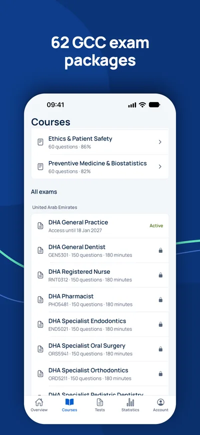
- هيئة الصحة بدبي – DHA Specialist Oral Surgery (ORS5941): 150 سؤال, 180 دقيقة, درجة النجاح 60 %
- هيئة الصحة بدبي – DHA Consultant Oral Surgery (COR6082): 100 سؤال, 180 دقيقة, درجة النجاح 70 %
Exodontia & impactions
Lesson 1Principles of exodontia: assessment, instruments and technique
Exodontia is the removal of teeth. At specialist level the examiner is not interested in whether you can use forceps; they want to know whether you can predict difficulty, choose the right technique before you start, and recognise when a simple extraction should become a surgical one. Most complications are decided in the planning phase, not in the chair.
Every extraction begins with three questions. Why is the tooth being removed (indication, and whether an alternative exists)? Is the patient fit for the procedure (medical history, drugs that affect bleeding or bone healing, previous radiotherapy)? And how difficult will this tooth be (crown, roots, bone, neighbouring structures)? A good periapical or panoramic radiograph answers most of the third question.
| Feature on assessment | Why it makes extraction harder |
|---|---|
| Heavily restored or root-filled crown | Brittle crown fractures under forceps; plan to section or raise a flap early |
| Divergent, curved or dilacerated roots | Roots cannot follow a single path of withdrawal and tend to fracture |
| Bulbous roots, hypercementosis | Root is wider apically than at the cervix; bone must be removed |
| Dense bone, older patient, ankylosis | Less elasticity for expansion of the socket |
| Sinus floor dipping between upper molar roots | Risk of oroantral communication or root displacement into the antrum |
| Isolated upper molar, especially last standing | Tuberosity is weakened by pneumatised sinus; risk of tuberosity fracture |
| Roots close to inferior alveolar canal or mental foramen | Risk of nerve injury |
Elevators move teeth using three simple mechanical principles. A wedge drives the blade between root and bone, which expands the socket and pushes the root out; a straight (Coupland or straight Warwick James) elevator used in the periodontal ligament space acts as a wedge. A lever pivots on a fulcrum (usually bone, never an adjacent tooth) so that small force on the handle produces large force at the tip; straight elevators also act as levers. The wheel and axle converts rotation of the handle into lifting at the tip; the cross-bar (Cryer, Winter) elevator is the classic example and is used to remove a remaining root once the other socket is empty, by engaging the inter-radicular bone.
Surgical (open) extraction is not a failure. It is the planned technique whenever crown or roots predict a fracture, and it is the rescue technique when forceps fail. The steps are the same for any tooth: adequate local anaesthesia, a full-thickness mucoperiosteal flap with a broad base and good blood supply, bone removal with a bur under saline irrigation, sectioning of the tooth so that each part has its own path of withdrawal, delivery, debridement of the socket, smoothing of sharp bone, irrigation and closure.
- Confirm consent, indication, side and tooth number; check medical history and anticoagulants.
- Give local anaesthesia and test it.
- Divide the periodontal ligament (luxator or periotome) to begin expansion with minimal trauma.
- Apply elevator or forceps; if no movement or crown is crumbling, convert to surgical approach rather than escalating force.
- Raise a full-thickness flap, remove buccal bone, section roots, and deliver each part separately.
- Inspect the tooth and socket, check for sinus communication in upper molars, irrigate, achieve haemostasis, suture.
- Give written postoperative instructions and analgesia plan.
Upper molars deserve special respect. In many adults the floor of the maxillary sinus dips between the roots. If you apply rotational force with forceps to divergent roots surrounded by a thin sinus floor, the whole block of bone can come away with the tooth, leaving a large oroantral communication. The safer technique is to section the crown so that the palatal and buccal roots are separated, then remove each root along its own path with gentle elevation. This keeps the sinus floor in place.
The isolated upper second or third molar is the classic setting for a tuberosity fracture. The tuberosity is weak because the sinus is large and there is no neighbouring tooth behind it. Signs are movement of the tooth together with the adjacent bone and a cracking sensation. If the fragment is still attached to the mucoperiosteum on both sides, it has a blood supply and can heal. Stop, free the tooth from the forceps, reposition the segment and splint it to adjacent teeth (for example with a wire-composite or arch-bar splint). The tooth is removed surgically after about 6 to 8 weeks once the bone has united, usually after the pulp is extirpated or the crown is reduced out of occlusion to relieve pain. If the fragment is small and stripped of soft tissue, it is dissected free and the soft tissue closed primarily.
Sources: Hupp JR, Ellis E, Tucker MR. Contemporary Oral and Maxillofacial Surgery, 7th ed. Elsevier, 2019 (chapters on principles of routine and complicated exodontia). · Fonseca RJ. Oral and Maxillofacial Surgery, 3rd ed. Elsevier, 2018. · Moore UJ. Principles of Oral and Maxillofacial Surgery, 6th ed. Wiley-Blackwell, 2011.
عن امتحان DHA / DOH / MOHAP Specialist Oral & maxillofacial surgery
كم عدد أسئلة امتحان DHA / DOH / MOHAP Specialist Oral & maxillofacial surgery؟+
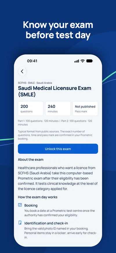
هيئة الصحة بدبي – DHA Specialist Oral Surgery (ORS5941): 150 سؤال, 180 دقيقة, درجة النجاح 60 %; هيئة الصحة بدبي – DHA Consultant Oral Surgery (COR6082): 100 سؤال, 180 دقيقة, درجة النجاح 70 %. يتم تأكيد العدد الدقيق للأسئلة والوقت ودرجة النجاح في حجزك.
ما المواضيع الأكثر وزنًا؟+
أكبر المواضيع هي Cysts & tumours (20 %), Exodontia & impactions (15 %), Infections (15 %). يغطي المقرر 6 موضوعًا و30 فصلًا.
كيف أستعد لامتحان DHA / DOH / MOHAP Specialist Oral & maxillofacial surgery؟+
ادرس الفصول موضوعًا تلو الآخر، وراجع الملاحظات المهمة، ثم تدرّب باختبارات تجريبية موقوتة بصيغة جهتك.
أسئلة
كم مدة الوصول؟+
يستمر الوصول 90 يومًا من الدفع للدولة والمهنة المختارتين.
ما الأجهزة التي يمكنني استخدامها؟+
أي متصفح على الحاسوب أو الجهاز اللوحي أو الهاتف، وتطبيق MediFlow بالحساب نفسه.
هل يمكنني استرداد المبلغ؟+
يرجى قراءة سياسة الاسترداد قبل الشراء. سياسة الاسترداد
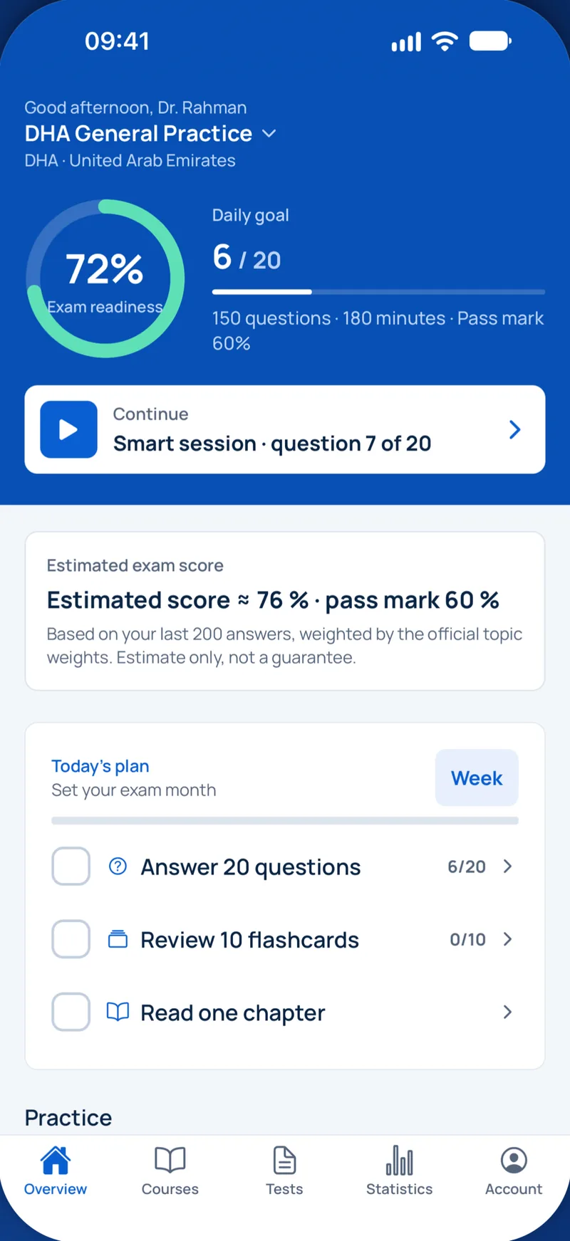
iPhone · iPad
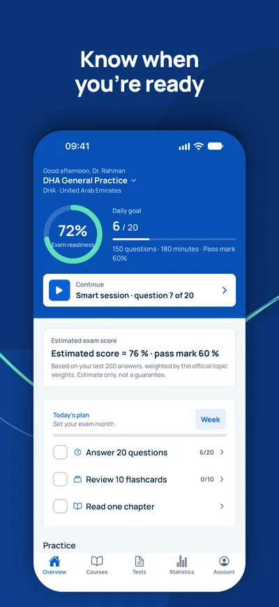
GCC Medical Exam Prep
استعدوا على iPhone لامتحانات الترخيص DHA وDOH وMOHAP وSCFHS وغيرها في دول الخليج.
هل الاختبار هو خطوتك المتبقية الوحيدة؟
تحقق أولًا مما إذا كنت تحتاج إلى الاختبار أصلًا وما المسمى المتوقع لك. يجيب المستشار عن أسئلتك مجانًا.
فحص الأهلية- 1استشارة مجانية عبر الفيديو أو الهاتف أو واتساب
- 2فحص الأهلية وفق المتطلبات الرسمية
- 3خطتك بأسعار ثابتة وخطوات واضحة