
🇴🇲 OMSB
Specialist Endodontics Oman
Specialist dentists preparing for the Endodontics exams of DHA, DOH, Qatar DHP and SCFHS

Pergunta de exemplo
A 25-year-old patient had an emergency pulpotomy on tooth 26 last week by another dentist and is referred to complete treatment. Which AAE pulpal diagnosis applies?
Keys: 1–5 or A–E to choose, Enter to check and continue, F to mark.
Original MediFlow content, not official exam questions. Medical review in progress.
Syllabus and high-yield notes
1. Diagnosis · 15 % peso no exame · 6 lessons+
- Always record two diagnoses: one pulpal and one apical, using the AAE terminology.
- Lingering pain after a thermal stimulus is the key feature of irreversible pulpitis; percussion tenderness defines symptomatic apical periodontitis, even with a vital pulp.
- Never start root canal treatment on the basis of a single test; reproduce the complaint and confirm with at least two findings.
- Radiolucencies next to teeth with normal sensibility are not endodontic until proven otherwise; think of cysts, dysplasia and tumours.
- Non-odontogenic pain (neuralgia, myofascial pain, sinusitis, cardiac pain) is suspected when dental tests are normal and local anaesthesia of the tooth gives no relief.
- CBCT is a problem-solving tool, not a routine test; metal and gutta-percha artefacts reduce its value for root fractures.
- Endo-periodontal lesions are classified first by root damage, then by periodontitis status and pocket grade (2017 classification).
- Cracks progress from craze line to split tooth; the prognosis depends on how far the crack extends apically.
- Diagnostic terminology: one pulpal and one apical diagnosis – Endodontic diagnosis answers two separate questions. First: what is the state of the pulp, the soft tissue inside the tooth? Second: what is the state of the apical tissues, meaning the periodontal ligament and bone around the root end? The American Association of Endodontists (AAE) built its terminology around these two questions, and specialist examiners expect every case to carry one pulpal and one apical diagnosis. The two are linked but not identical. A vital but inflamed pulp can already make the periodontal ligament tender, and a necrotic pulp can sit for years with healthy-looking apical bone.
- Clinical examination and pulp testing – A good endodontic diagnosis is built like a court case. The history gives the complaint, the clinical tests try to reproduce it, and the radiograph adds supporting evidence. A safe rule taught by Cohen's Pathways of the Pulp is that treatment should never start on the basis of one test alone. You should reproduce the patient's complaint and have at least two findings that point to the same tooth and the same diagnosis. Compare with control teeth every time: the contralateral tooth and an adjacent healthy tooth tell you what a 'normal' response looks like for this patient.
- Radiographic diagnosis, CBCT and lesions that are not endodontic – Radiographs support a diagnosis; they rarely make it alone. A periapical radiograph taken with a paralleling technique and a film holder is the standard starting image. A second view with a mesial or distal horizontal angle separates superimposed roots (buccal object rule: the object farther from the film, the buccal one, moves in the opposite direction to the tube shift; the lingual object moves with the tube, remembered as SLOB, same lingual opposite buccal).
- Special diagnostic problems: cracks, endo-perio lesions, trauma and non-odontogenic pain – Some diagnoses fail not because the tests are wrong but because the problem lies outside the usual pulp-to-apex pathway. This chapter covers four groups that specialist examiners like: cracked teeth, combined endodontic-periodontal lesions, teeth after trauma, and pain that comes from outside the teeth.
- Patient safety, ethics and professionalism in endodontics – Endodontics is done in a small, dark space with sharp instruments, caustic irrigants and ionising radiation, often on anxious patients who cannot see what is happening. Most harm in the endodontic chair is preventable. The World Health Organization Patient Safety Curriculum Guide (2011) frames safety as a system property: errors are expected, and good systems catch them before they reach the patient. This chapter applies that thinking to consent, wrong-site prevention, the airway, irrigants, radiation, records, referral and professional conduct.
- Medically compromised patients: endodontic decisions and precautions – Many patients who need root canal treatment take medicines or have conditions that change risk. Endodontics is often the safer choice in these patients because it avoids extraction, a wound in bone. This chapter covers the conditions that examiners ask about most: antiresorptive and antiangiogenic drugs, anticoagulants, head and neck radiotherapy, infective endocarditis risk, diabetes, cardiovascular disease, pregnancy, immunosuppression and bleeding disorders. The principle throughout: take a full medical history, talk to the patient's physician when needed, and adapt the plan rather than avoid care.
2. Anatomy & access · 15 % peso no exame · 5 lessons+
- Know the common extra canals: MB2 in maxillary molars, second canal in mandibular incisors and premolars, distolingual canal or radix in mandibular molars.
- Krasner and Rankow laws (centrality, concentricity, colour change, symmetry, orifice location) guide the search for orifices.
- Straight-line access means removing obstructions such as dentine shelves, not enlarging the whole cavity.
- A canal that suddenly disappears on a radiograph usually splits; use angled views or CBCT.
- The apical foramen often lies short of and lateral to the radiographic apex; apex locators are more accurate than radiographs for length.
- In calcified teeth use magnification, ultrasonics and, for anterior teeth, guided endodontics to avoid perforations.
- Danger zones: mesial concavity of maxillary first premolars and the distal wall of mesial roots in mandibular molars.
- Dens invaginatus is classified by Oehlers; type III cases often need CBCT and separate treatment of the invagination.
- The pulp space: canal configurations and apical anatomy – Endodontic treatment fails most often where the clinician did not know the anatomy: a missed canal, an uncleaned isthmus, a file that stopped short of or passed beyond the real end of the canal. This chapter builds the mental model of the pulp space that every later decision depends on. Think of the pulp space as a system, not a tube. It has a chamber in the crown, one or more root canals, and many small side branches that connect the pulp to the periodontal ligament (PDL).
- Tooth by tooth: roots, canals and the extra canal you must look for – Most missed canals are not rare oddities. They are common variants that the operator did not actively search for. The rule is simple: for every tooth, know the usual number of canals, the most frequent extra canal and where its orifice lies. Prevalence figures vary widely between studies and ethnic groups, so learn ranges and patterns rather than single numbers.
- Access cavity design: orifice laws, straight-line access and restorations – The access cavity is the first and least forgiving step of root canal treatment. Its aims are to remove the whole pulp chamber roof, find every orifice, give instruments a straight path into the coronal and middle thirds of each canal, and preserve as much sound dentine as possible. A poor access causes missed canals, ledges, instrument separation and perforations, problems that no later step can fully correct.
- Difficult anatomy: calcification, developmental anomalies and 3D imaging – Some teeth have anatomy that defeats routine technique. The specialist must recognise these situations before starting, choose the right imaging, and use magnification, ultrasonics and sometimes navigation. The key principle is to remove dentine only where you can see or have planned, because perforation in these teeth is common and often ruins the prognosis.
- Landmark studies of root canal anatomy and what they still teach – Almost everything we say about canal anatomy comes from a small number of studies that cleared, sectioned or scanned extracted teeth. Specialist examinations treat these studies as core literature: you are expected to know who described what, how the work was done, and where its limits lie. This chapter walks through the classic anatomy papers in historical order, explains the methods behind them, and shows how newer micro-CT and CBCT work has changed the numbers. The aim is not to memorise percentages to the decimal point, but to understand why a percentage from one study may not fit the patient in front of you.
3. Treatment · 25 % peso no exame · 5 lessons+
- Sodium hypochlorite dissolves organic tissue and kills bacteria; EDTA removes the inorganic smear layer. Use them in sequence, never mixed.
- Single- and multiple-visit treatment give similar healing; choose by case complexity and time.
- Antibiotics are an adjunct only for systemic involvement or spreading infection; drainage and canal debridement come first.
- For pain, combine ibuprofen and paracetamol; opioids and antibiotics are not first line.
- The presence and size of a periapical lesion is the strongest preoperative prognostic factor; quality of the root filling and coronal seal are key intraoperative factors.
- Posterior root-filled teeth need cuspal coverage; posts retain cores and do not strengthen roots.
- A separated instrument in a vital case without apical periodontitis has little effect on the outcome; weigh removal risks carefully.
- Review healing at 1 year and up to 4 years before declaring failure.
- Shaping and Disinfecting the Root Canal System – Root canal treatment has one biological aim: to prevent or cure apical periodontitis. Apical periodontitis is inflammation of the tissues around the root end, caused by microorganisms living in the root canal system. Everything we do mechanically and chemically serves that aim. Files create space and shape; irrigants do most of the actual disinfection; the filling and the coronal restoration stop bacteria from coming back. If you keep this logic in mind, most exam questions on technique answer themselves.
- Obturation, Endodontic Emergencies, Pain and Antibiotics – Obturation fills the shaped and disinfected canal to entomb remaining bacteria, prevent fluid from feeding them and block reinfection. It does not disinfect. A good filling is dense, has no voids, ends close to working length and follows the shape that was prepared. Gutta-percha is the core material; a sealer fills the gaps between gutta-percha and dentine and between gutta-percha cones.
- Procedural Errors and Non-Surgical Retreatment – Procedural errors are mishaps during treatment: ledges, transportation, perforations, separated instruments and overextension. Their importance is not the error itself but whether it prevents disinfection of the infected part of the canal. A mishap in a vital tooth, where the apical canal is usually not infected, matters far less than the same mishap in a necrotic tooth with apical periodontitis. This principle, from outcome studies such as those by Spili and colleagues and by Ng and colleagues, guides almost every decision.
- Restoring the Root-Filled Tooth and Judging the Outcome – A root canal treatment is only as good as the restoration that follows it. Coronal leakage, the entry of saliva and bacteria between the restoration and the tooth, can reinfect a well-filled canal within weeks. Root-filled teeth also fail by fracture. The main reason is not that the dentine becomes brittle after the pulp is removed; it is the loss of tooth structure from caries, previous restorations and access, especially loss of the marginal ridges. Restoration planning therefore aims to seal the canal and protect what remains.
- Endodontic mishaps: hypochlorite accidents, perforations, nerve injury and emphysema – An endodontic mishap is an unwanted event during treatment that can harm the patient or the prognosis of the tooth. Ledges, transportation and separated instruments are covered in the chapter on procedural errors. This chapter deals with the mishaps that cause acute harm beyond the tooth: sodium hypochlorite extrusion, perforations of the root or pulp floor, overextension of materials into the mandibular canal or sinus, air emphysema and the loss of instruments into the airway. For each, know the cause, the warning signs, the immediate response and the prognosis.
4. Vital pulp therapy & regeneration · 15 % peso no exame · 5 lessons+
- In deep caries without irreversible pulpitis, avoid exposure with selective caries removal in one session.
- Full pulpotomy with calcium silicate is an accepted alternative to root canal treatment in mature teeth with irreversible pulpitis signs.
- Direct visual assessment and control of bleeding within about 5 minutes are better guides than symptoms.
- Calcium silicate cements give more predictable dentine bridges than calcium hydroxide.
- Bismuth oxide in MTA can stain; use zirconium-based materials in aesthetic zones.
- Regenerative endodontic procedures use low concentration hypochlorite, EDTA, no vasoconstrictor at the second visit and a calcium silicate barrier.
- The primary goal of REP is healing; root thickening and sensibility are secondary and tertiary goals.
- Follow vital pulp therapy for up to 4 years because failure can be silent.
- Pulp biology and the logic of keeping the pulp vital – Vital pulp therapy (VPT) means every treatment that aims to keep all or part of the dental pulp alive and healthy instead of removing it. The idea is simple. A living pulp defends the tooth, keeps sensation, and in a young patient continues to build the root. A root-filled tooth has none of these abilities and is more likely to fracture or be lost later. Modern guidelines from the European Society of Endodontology (ESE, 2019) and the American Association of Endodontists (AAE, 2021) therefore push the profession towards preserving pulp whenever the tissue can heal.
- Deep caries management: selective removal and stepwise excavation – Deep caries is a lesion that reaches the inner quarter of the dentine on a radiograph, with a zone of hard or firm dentine still separating it from the pulp. Extremely deep caries penetrates the entire thickness of dentine so that exposure is certain. The ESE uses this distinction because the management differs. In deep caries with a vital pulp and no signs of irreversible pulpitis, the main aim is to avoid exposure. An exposed pulp in a carious field is contaminated, and the outcome of capping in such a situation is less predictable than leaving a thin layer of affected dentine in place.
- Pulp capping and pulpotomy: technique and materials – When the pulp is exposed, by caries, trauma or during cavity preparation, the clinician chooses between direct pulp capping, partial pulpotomy and full pulpotomy. The choice depends on the size and cause of the exposure, the appearance of the tissue and whether bleeding stops. All three share the same principles: rubber dam, magnification, a disinfecting solution to clean the wound, a bioactive capping material, and an immediate tight restoration.
- Immature necrotic teeth: regeneration, apexification and follow-up – When an immature permanent tooth loses its pulp, usually after trauma or caries in a child, root development stops. The canal stays wide, the walls thin and the apex open. Such a tooth is hard to clean and fill conventionally and is prone to cervical fracture. Two strategies exist: apexification, which creates an apical barrier, and regenerative endodontic procedures (REP, also called revitalisation), which aim to bring vital tissue back into the canal.
- Classic and current literature in pulp biology and vital pulp therapy – A fifth of a specialist endodontic examination may draw on literature. In the field of pulp biology and vital pulp therapy, a handful of studies explain why we practise as we do today. This chapter presents them as a story: first the proof that bacteria cause pulp and periapical disease, then the discovery that clinical symptoms predict pulp histology poorly, then the clinical trials that moved practice from complete caries removal and root canal treatment toward minimally invasive, pulp-preserving care. For each study, know the question, the design and the main finding.
5. Surgery · 15 % peso no exame · 5 lessons+
- Microsurgery has a much higher success rate than traditional surgery.
- Resect 3 mm of the apex with a minimal bevel.
- Use ultrasonic tips to prepare a 3 mm root-end cavity and fill with a calcium silicate material.
- Flap design preserves papillae in aesthetic areas; protect the mental nerve.
- Remove ferric sulphate before closure.
- Intentional replantation is an option when surgical access is very difficult.
- Healing is classified as complete, incomplete, uncertain or unsatisfactory.
- Send tissue for histopathology to confirm the diagnosis.
- Endodontic surgery today: indications, case selection and outcomes – Endodontic surgery (also called periradicular or apical surgery) treats a tooth whose periapical disease cannot be resolved, or should not be resolved, through the crown. The surgeon reaches the root tip through the bone, removes the diseased tissue and the infected apex, and seals the canal from the root end. It is not a replacement for good root canal treatment; it is the next step when the orthograde route is exhausted or blocked.
- Haemostasis, flap design and access: working safely around anatomy – Good surgery starts before the incision. Two goals guide this chapter: a dry, clear field, and a flap that gives access without damaging gingiva or nerves.
- Resection, inspection, root-end preparation and filling – This chapter covers the core of the operation: resect the apex, inspect the cut surface, prepare a root-end cavity and seal it. The numbers here are classic examination material, and each has a reason.
- Closure, complications, alternatives and judging healing – The last chapter brings the operation to a close and deals with what comes after: suturing and aftercare, complications, alternatives to conventional apical surgery, and judging whether the tooth has healed.
- Outcome literature: retreatment, surgery, implants and working with other specialties – Specialist examinations ask not only how to do endodontic surgery but whether to do it, compared with nonsurgical retreatment, extraction with an implant, or no treatment. Those choices rest on outcome literature. This chapter explains how outcome is measured, summarises the classic and current studies on primary treatment, retreatment, surgery and implants, and closes with the role of the endodontist alongside periodontists, prosthodontists, orthodontists and oral surgeons.
6. Trauma · 15 % peso no exame · 5 lessons+
- Replant avulsed permanent teeth whenever possible.
- Use a flexible splint; know splint times by injury type.
- Start root canal treatment within 2 weeks in mature avulsed or intruded teeth.
- Immature teeth can revascularise; monitor them.
- Root fractures in the cervical third need a long splint period.
- Treat infection-related resorption immediately.
- Decoronation preserves bone in growing patients with ankylosis.
- Use amoxicillin rather than tetracyclines in children under 12.
- Examination, classification and fractures of permanent teeth – Traumatic dental injuries are common in children, adolescents and young adults, and the upper central incisors are hit most often. The outcome of many injuries is decided in the first hour, so a specialist must be able to classify the injury quickly, choose the correct emergency treatment and plan follow-up. The reference standard worldwide, and the basis of most licensing exam questions, is the set of guidelines published by the International Association of Dental Traumatology (IADT) in 2020. This chapter covers the examination and the fractures of the hard tissues; the next chapters cover luxations, avulsion and the late complications.
- Luxation injuries and splinting – A luxation injury is any injury in which the tooth is loosened or displaced within its socket. Six types are described, ordered roughly by increasing damage: concussion, subluxation, extrusion, lateral luxation, intrusion and avulsion (complete displacement, treated in the next chapter). The more the tooth moves, the more the neurovascular bundle at the apex and the PDL are damaged. Knowing the definitions exactly is essential, because many exam items describe a clinical picture and ask for the diagnosis.
- Avulsion of permanent teeth – Avulsion means the tooth has come completely out of its socket. It is the most serious dental injury because the PDL cells left on the root surface start to die as soon as they dry out. The prognosis depends mainly on what happens in the first hour: how long the tooth stayed dry, in what medium it was stored, and how quickly it was replanted. The IADT states that permanent teeth should be replanted whenever possible, even after long dry times, because a replanted tooth keeps the alveolar bone, height and width for some years, which keeps future options open, especially in growing patients.
- Healing complications, resorption and injuries in the primary dentition – Most late problems after dental trauma arise either in the pulp (necrosis, infection, canal obliteration) or at the root surface (resorption, ankylosis). Long-term follow-up for at least 5 years exists to detect these early, because several of them, if treated in time, can be stopped. This chapter explains the types of root resorption, how to manage ankylosis in a growing patient, and how injuries to primary teeth differ.
- Long-term care after trauma: ankylosis in growing patients, decoronation, autotransplantation and teamwork – The first visit after a dental injury decides much, but many traumatised teeth create their real problems months or years later. Replacement resorption, infraposition, crown discolouration and tooth loss in a growing child require plans that span the whole of adolescence and need orthodontists, paediatric dentists and prosthodontists. This chapter covers follow-up schedules, the ankylosed incisor in a growing patient, decoronation, autotransplantation, orthodontic movement of traumatised teeth and the literature behind these decisions.
Diagnosis
Lesson 1Diagnostic terminology: one pulpal and one apical diagnosis
Endodontic diagnosis answers two separate questions. First: what is the state of the pulp, the soft tissue inside the tooth? Second: what is the state of the apical tissues, meaning the periodontal ligament and bone around the root end? The American Association of Endodontists (AAE) built its terminology around these two questions, and specialist examiners expect every case to carry one pulpal and one apical diagnosis. The two are linked but not identical. A vital but inflamed pulp can already make the periodontal ligament tender, and a necrotic pulp can sit for years with healthy-looking apical bone.
Why insist on two labels? Because they lead to different decisions. The pulpal diagnosis decides whether the pulp can be kept (vital pulp therapy, a new restoration) or must be removed (root canal treatment or extraction). The apical diagnosis tells you whether infection has spread beyond the root, whether drainage or antibiotics might be needed, and what radiographic healing you should expect at follow-up.
| AAE pulpal diagnosis | Key findings | Usual consequence |
|---|---|---|
| Normal pulp | No symptoms; normal, short response to cold that stops when the stimulus is removed | No endodontic treatment |
| Reversible pulpitis | Sharp, short pain to cold or sweets that stops within seconds after the stimulus is removed; cause usually caries, exposed dentine or a new restoration | Remove the cause; pulp expected to recover |
| Symptomatic irreversible pulpitis | Lingering pain after thermal stimulus (often more than 30 seconds), spontaneous pain, referred pain, pain on lying down | Pulpotomy or pulpectomy (root canal treatment), or extraction |
| Asymptomatic irreversible pulpitis | No symptoms, but a vital pulp that cannot heal, e.g. deep caries that will expose the pulp on removal | As for symptomatic irreversible pulpitis |
| Pulp necrosis | No response to sensibility tests; usually no pulpal pain | Root canal treatment or extraction |
| Previously treated | Tooth already root filled (other than intracanal medicament) | Assess quality and apical status; retreatment, surgery or monitoring |
| Previously initiated therapy | Partial treatment already done, e.g. pulpotomy or pulpectomy started | Complete the treatment |
The single most useful sign for separating reversible from irreversible pulpitis is how long the pain lasts after the stimulus is taken away. In reversible pulpitis the A-delta nerve fibres in the dentine fire briefly and then stop. In irreversible pulpitis the inflamed pulp has raised tissue pressure and sensitised C fibres; the pain is dull, throbbing and lingers. Spontaneous pain without any trigger also points to irreversible disease. Dentine hypersensitivity looks similar to reversible pulpitis: short sharp pain to air or cold on exposed cervical dentine that stops immediately. It is explained by fluid movement in open dentinal tubules (hydrodynamic theory) and is not pulp disease at all.
| AAE apical diagnosis | Key findings |
|---|---|
| Normal apical tissues | Not tender to percussion or palpation; intact lamina dura and uniform PDL space |
| Symptomatic apical periodontitis | Pain on biting, percussion or palpation; may or may not have a radiographic lesion; the pulp may still be vital |
| Asymptomatic apical periodontitis | Apical radiolucency without clinical symptoms |
| Acute apical abscess | Rapid onset, spontaneous pain, tenderness, pus formation and swelling; possible fever, malaise, lymphadenopathy |
| Chronic apical abscess | Gradual onset, little or no discomfort, intermittent pus discharge through a sinus tract |
| Condensing osteitis | Diffuse radiopaque bone around the apex, a reaction to low-grade inflammation, usually from the pulp |
Three points in this table are tested again and again. First, symptomatic apical periodontitis is defined by tenderness, not by the radiograph and not by pulp status. A tooth with irreversible pulpitis whose inflammation has reached the ligament is tender to percussion, so it gets both labels. Second, the difference between symptomatic apical periodontitis and an acute apical abscess is swelling, meaning pus collecting in the tissues. Pain alone does not make an abscess. Third, a chronic apical abscess is defined by the sinus tract. A sinus tract usually means a necrotic pulp, and it is the pathway that lets the abscess drain, which is why the patient often feels little.
Condensing osteitis deserves a separate word. It appears as a diffuse radiopacity that blends into the surrounding bone around the apex of a tooth with an inflamed or necrotic pulp, most often a mandibular molar. The bone reacts to mild, long-standing irritation by laying down more trabeculae. It differs from idiopathic osteosclerosis (dense bone island), which sits next to a tooth with a normal pulp and does not need treatment. After successful root canal treatment, condensing osteitis may partly regress.
Old literature and some textbooks use the term 'phoenix abscess' for an acute flare-up of a previously chronic apical lesion. The current AAE glossary treats this as an acute exacerbation of asymptomatic apical periodontitis. When a tooth with a known silent periapical radiolucency suddenly becomes painful, think of this term.
Sources: American Association of Endodontists. Glossary of Endodontic Terms, 10th ed. 2020. · AAE Consensus Conference Recommended Diagnostic Terminology. J Endod 2009;35:1634. · Berman LH, Hargreaves KM (eds). Cohen's Pathways of the Pulp, 12th ed. Elsevier 2020, chapter on diagnosis. · Torabinejad M, Fouad AF, Shabahang S. Endodontics: Principles and Practice, 6th ed. Elsevier 2020.
Sobre o exame OMSB Specialist Endodontics
Quantas questões tem o exame OMSB Specialist Endodontics?+
não publicado. O número exato de questões, o tempo e a nota mínima são confirmados na sua marcação.
Quais temas têm mais peso?+
Os maiores temas são Treatment (25 %), Diagnosis (15 %), Anatomy & access (15 %). O curso abrange 6 temas com 31 capítulos no total.
Como me preparo para o exame OMSB Specialist Endodontics?+
Estude os capítulos tema a tema, reveja as notas essenciais e pratique com simulados cronometrados no formato da sua autoridade.
Perguntas
Quanto tempo tenho acesso?+
O acesso dura 90 dias a partir do pagamento para o país e a profissão escolhidos.
Que dispositivos posso usar?+
Qualquer navegador no computador, tablet ou telemóvel, e a app MediFlow com a mesma conta.
Posso pedir reembolso?+
Leia a nossa política de reembolso antes de comprar. Política de reembolso

iPhone · iPad
GCC Medical Exam Prep
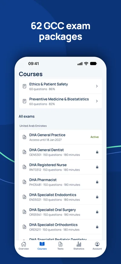
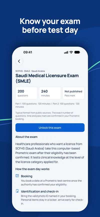
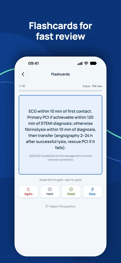
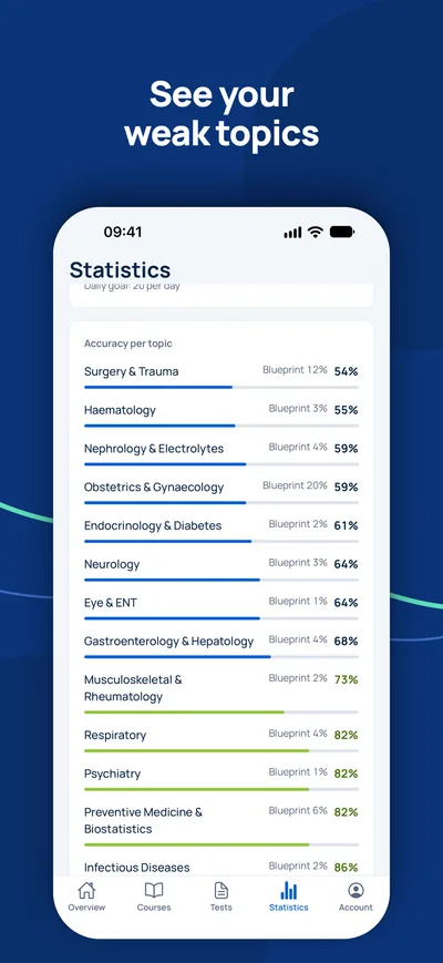
Prepare-se no seu iPhone para DHA, DOH, MOHAP, SCFHS e outros exames de licença do Golfo.

O exame é o seu único passo em aberto?
Verifique primeiro se precisa do exame e que título pode esperar. Um consultor responde às suas perguntas gratuitamente.
Verificação de elegibilidade- 1Consulta gratuita por vídeo, telefone ou WhatsApp
- 2Verificação de elegibilidade segundo os requisitos oficiais
- 3O seu plano com preços fixos e próximos passos claros