
🇦🇪 DHA · DOH · MOHAP
Dentistry United Arab Emirates
General dentists preparing for GCC licensing exams
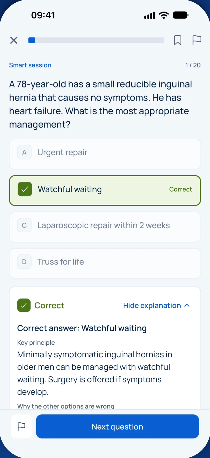

Sample question
Below which approximate plaque-fluid pH (critical pH) does hydroxyapatite of sound enamel begin to dissolve under typical oral conditions?
Keys: 1–5 or A–E to choose, Enter to check and continue, F to mark.
Original MediFlow content, not official exam questions. Medical review in progress.
Syllabus and high-yield notes
1. Operative dentistry & cariology · 9 % exam weight · 5 lessons+
- Caries is a dynamic, biofilm-mediated, sugar-driven disease. Enamel demineralises when plaque pH falls below about 5.5 and root dentine already at about 6.2–6.7. Fluorapatite-like mineral only dissolves at about pH 4.5.
- Fluoride acts mainly topically: low, constant levels in plaque fluid and saliva inhibit demineralisation and promote remineralisation. Know the concentrations: toothpaste 1000–1500 ppm, prescription toothpaste 5000 ppm (1.1% NaF), 1.23% APF gel 12,300 ppm, 5% NaF varnish 22,600 ppm, 38% SDF 44,800 ppm. The probably toxic dose is 5 mg F/kg body weight.
- ICDAS: 0 sound; 1 first visual change, seen only after air-drying; 2 distinct visual change, seen wet; 3 localised enamel breakdown; 4 underlying dark shadow from dentine; 5 distinct cavity with visible dentine; 6 extensive cavity involving more than half the surface. The ADA 2023 guideline calls ICDAS 3–4 moderate and ICDAS 5–6 advanced lesions.
- Treat non-cavitated lesions without drilling (ADA 2018). Use sealants (with or without varnish) on occlusal surfaces. On proximal lesions that radiographs show limited to enamel or the outer third of dentine, use varnish, resin infiltration or sealant. For root lesions, 5000 ppm toothpaste comes first. When restoration is not feasible, 38% SDF arrests cavitated coronal lesions.
- G.V. Black: Class I covers pits and fissures, including the lingual pits of maxillary incisors and the buccal/lingual pits of molars. Class II is proximal posterior, Class III proximal anterior without the incisal angle, Class IV proximal anterior including the incisal angle, and Class V the gingival third of facial or lingual surfaces. Class VI (added later) covers incisal edges and cusp tips.
- Etch-and-rinse bonding: 30–40% phosphoric acid for about 15 s, rinse, leave the dentine visibly moist (wet bonding), then apply primer and bond. Over-drying collapses the collagen network. Saliva on etched enamel calls for rinsing and re-etching. With mild self-etch adhesives, etch uncut enamel margins selectively with phosphoric acid.
- Composite shrinkage stress rises with the C-factor (bonded/unbonded surfaces). A Class I cavity, at about 5, is the worst. Place increments of 2 mm or less (unless the material is a bulk-fill), keep the light guide as close as possible, and use a pre-contoured sectional matrix with a separation ring for tight proximal contacts.
- Amalgam preparation needs a 90° butt-joint cavosurface margin, rounded internal line angles and a pulpal depth of about 1.5–2 mm. Cap a cusp when the outline reaches two-thirds of the distance from the primary groove to the cusp tip: reduce it about 1.5 mm (non-functional cusp) or 2 mm (functional cusp).
- Deep caries in a vital tooth without signs of irreversible pulpitis: remove carious tissue selectively, leaving soft or firm dentine over the pulp and hard dentine at the periphery, then seal in one visit (ADA 2023, ESE 2019). For a small exposure in an asymptomatic tooth, achieve haemostasis with NaOCl and cap with a hydraulic calcium silicate cement.
- Non-carious tooth wear: erosion is chemical (cupping, palatal incisor surfaces with intrinsic acid), attrition comes from tooth-to-tooth contact (matching flat facets), abrasion from a foreign body such as a toothbrush (cervical notches), and abfraction is a disputed load-related wedge-shaped lesion. Record it with BEWE. For localised anterior wear, the Dahl concept creates space without tooth reduction.
- Restorations fail mainly through secondary caries and fracture. Repairing a localised defect is preferred to replacement because it preserves tooth structure. Glass-ionomer or resin-modified glass-ionomer is suggested for root caries and helps in high-risk patients with limited moisture control.
- Caries as a disease: biofilm, critical pH and fluoride – Modern cariology no longer treats caries as a hole that must be drilled. Caries is a dynamic, biofilm-mediated, sugar-driven disease that causes a net loss of mineral from tooth tissue. The hole (the cavity) is only the late sign of a process that has been running for months or years. Understanding the process explains almost every management decision you will meet in the exam: why we seal, why we use fluoride, why we leave soft dentine over the pulp, and why diet frequency matters.
- Detecting, classifying and managing lesions without drilling – Before deciding on treatment you need two answers: how far has the lesion progressed, and is it active? Detection is visual and tactile on clean, dry teeth, supported by bitewing radiographs for proximal surfaces. A blunt probe (such as the ball-ended WHO probe) is used gently to feel surface texture; forcing a sharp explorer into a fissure is outdated because it can create a cavity.
- Instruments, isolation and the amalgam preparation – When a lesion is cavitated and cannot be cleaned or arrested, it needs a restoration. The preparation design depends on the material. Amalgam needs a mechanically retentive shape with bulk; composite relies on adhesion and allows a minimal, lesion-driven outline. This chapter covers the tools, the isolation and the classic amalgam principles that examiners still test.
- Adhesion and resin composite placement – Composite restorations depend entirely on adhesion. A well-bonded composite allows a small, lesion-driven preparation and reinforces the remaining tooth, but bonding is technique-sensitive. Most exam questions in this area test whether you understand why each step is done.
- Deep caries, the pulp, tooth wear and failing restorations – This chapter covers the clinical decisions that carry the most risk: how much carious dentine to remove near the pulp, what to do when the pulp is exposed, how to recognise non-carious tooth wear, and when to repair rather than replace a restoration.
2. Dental materials · 1 % exam weight · 5 lessons+
- Stress–strain curve: the slope of the linear part is the modulus of elasticity (stiffness). The area under the elastic part up to the proportional limit is resilience; the total area to fracture is toughness. Brittle materials (ceramics, amalgam, gypsum) are far weaker in tension than in compression. Knoop or Vickers tests measure the hardness of brittle materials such as enamel and porcelain.
- Alginate sets by a double-decomposition reaction between soluble alginate and calcium sulfate, delayed by trisodium phosphate. Warmer water speeds setting and cold water slows it. The gel loses water (syneresis) or absorbs water (imbibition), so pour promptly.
- Elastomers: polysulfide (lead dioxide, water by-product, highest tear strength), condensation silicone (ethanol by-product, more shrinkage), addition silicone (platinum catalyst, most dimensionally stable, releases hydrogen gas, inhibited by sulfur from latex) and polyether (hydrophilic, very stiff, swells in water). Being viscoelastic, all should be removed with a quick snap.
- Gypsum: plaster (beta-hemihydrate) needs more mixing water than stone and die stone (alpha-hemihydrate). Approximate water/powder ratios: model plaster 0.45–0.50, dental stone 0.28–0.30, die stone 0.22–0.24. More water gives a slower set, lower strength and less expansion. Potassium sulfate and terra alba accelerate setting; borax and citrates retard it.
- Cements: zinc phosphate sets exothermically and is very acidic at insertion; mix it on a cool slab in increments. Polycarboxylate was the first cement to bond chemically. Glass-ionomer sets by an acid–base reaction (calcium then aluminium polyacrylate), releases fluoride and must be protected from early water contact and drying out. Eugenol inhibits resin polymerisation. Luting film thickness should not exceed 25 µm.
- Resin composites: Bis-GMA/UDMA diluted with TEGDMA, silanated filler, camphorquinone plus amine (absorption peak about 468 nm). Volumetric shrinkage falls as the filler volume fraction rises. Microfills polish best but are weaker; hybrids and nanohybrids are all-purpose. Alternative photoinitiators (TPO, Ivocerin) need violet light from a polywave LED.
- Amalgam: in low-copper alloys the gamma-2 phase (Sn7–8Hg) is the weakest, most corrosion-prone phase and drives creep and marginal breakdown. High-copper alloys replace it with Cu6Sn5 (eta-prime). Zinc-containing alloys contaminated with moisture expand days later as hydrogen forms. Dissimilar metals in contact cause galvanic pain.
- Ceramics: strength rises from feldspathic porcelain (most aesthetic, weakest) through leucite-reinforced and lithium disilicate glass-ceramics to zirconia. Glass-ceramics are etched with hydrofluoric acid and silanated. Zirconia has no glass phase; it is air-abraded at low pressure and bonded with MDP. Zirconia is toughened by a tetragonal-to-monoclinic transformation with about 4% volume expansion.
- Alloys: a high-noble alloy contains at least 60 wt% noble metal, including at least 40 wt% gold; a noble alloy at least 25 wt% noble metal; a predominantly base alloy less than 25 wt%. Co–Cr is about twice as stiff as gold and much lighter, so clasps engage shallower undercuts. Nickel is the most common metal allergen. The metal's CTE is set slightly higher than the porcelain's so the porcelain ends up in compression.
- Investment and casting: gypsum-bonded investments are limited to about 700 °C (gold alloys). Phosphate-bonded investments are used for metal-ceramic and base-metal alloys, and silica-bonded investments mainly for Co–Cr frameworks. Setting, hygroscopic and thermal expansion of the investment compensate for the alloy's solidification shrinkage. Sprue diameter, a reservoir, venting and the burnout schedule prevent porosity and fins.
- PMMA denture base: the powder contains PMMA beads and the initiator benzoyl peroxide; the liquid contains MMA, the inhibitor hydroquinone and a cross-linker. Pack at the dough stage. Rapid heating boils monomer in thick sections (gaseous porosity); too little dough or pressure gives contraction porosity. Self-cure resins (tertiary-amine activator) leave more residual monomer, which can irritate mucosa and sensitise.
- Material properties: stress, strain, hardness and biocompatibility – Every material question becomes easier once you can read a stress-strain curve. Stress is the internal force per unit area that resists an applied load (measured in megapascals, MPa). Strain is the change in length divided by the original length, so it has no unit. If you load a test bar and plot stress against strain, the shape of the curve tells you how stiff, strong, elastic and tough the material is.
- Impression materials and gypsum products – An impression is a negative copy of the teeth and tissues; the cast or die poured into it is the positive. Accuracy is lost at every step, so you need to know how each material sets, how it can distort and how it interacts with gypsum.
- Dental cements and resin composites – Cements are used to lute indirect restorations, as liners and bases, and as restorative materials. They are classified by their chemistry, and the chemistry explains their handling. Most water-based cements are acid-base reactions between a basic powder and an acidic liquid.
- Amalgam, casting alloys and investments – Dental amalgam is made by mixing liquid mercury with an alloy powder of silver, tin and copper, sometimes with zinc. The powder particles can be lathe-cut (irregular filings), spherical or a blend (admixed). Spherical alloys need less mercury and less condensation force; lathe-cut alloys need firmer condensation but make it easier to establish proximal contacts.
- Dental ceramics and denture base resins – Dental ceramics range from glassy, translucent porcelains to dense polycrystalline zirconia. The general rule is simple: the more glass, the more translucent and aesthetic the material, but the weaker it is, and the more it can be etched for adhesive bonding. The more crystalline, the stronger and more opaque, and the less etchable.
3. Endodontics · 12 % exam weight · 6 lessons+
- Always give two diagnoses: one pulpal (normal pulp, reversible pulpitis, symptomatic or asymptomatic irreversible pulpitis, pulp necrosis, previously treated, previously initiated therapy) and one apical (normal apical tissues, symptomatic or asymptomatic apical periodontitis, acute or chronic apical abscess, condensing osteitis). Lingering thermal pain and spontaneous pain point to irreversible pulpitis; a sinus tract with little discomfort points to a chronic apical abscess.
- Cold, heat and electric tests are sensibility tests: they show nerve response, not blood supply. After trauma and in immature teeth they are often falsely negative, so do not start root canal treatment on a negative test alone. Pulse oximetry and laser Doppler flowmetry measure blood flow.
- Expect extra canals: MB2 in more than half of maxillary first molars (palatal to MB1), a lingual canal in many mandibular incisors, C-shaped systems in mandibular second molars (especially East Asian patients) and a distolingual radix entomolaris in mandibular first molars. Angled radiographs (SLOB rule), magnification and CBCT help to find them.
- Working length should end at the apical constriction, about 0.5–1 mm short of the radiographic apex. Combine an electronic apex locator with a radiograph; the apex locator's 'apex' or '0.0' reading marks the major foramen, not the working length.
- Irrigation: sodium hypochlorite (0.5–6%) dissolves organic tissue and kills bacteria; 17% EDTA for about 1 minute removes the inorganic smear layer; chlorhexidine 2% is substantive but does not dissolve tissue. Never let hypochlorite meet chlorhexidine (para-chloroaniline precipitate); use a passive, side-vented needle 1–3 mm short of working length to avoid extrusion.
- Rotary NiTi files fail by cyclic (flexural) fatigue in curved canals without warning, or by torsional overload when the tip binds, which often shows unwinding. A reproducible glide path, limited reuse, heat-treated alloys and avoiding prolonged rotation at one level reduce separation.
- Vital pulp therapy is preferred whenever the pulp can be kept: hydraulic calcium silicate cements (MTA, Biodentine) are the materials of choice for direct capping and pulpotomy. In traumatic exposures of young teeth, a partial (Cvek) pulpotomy is the standard treatment.
- For a necrotic immature tooth, a regenerative endodontic procedure (disinfection, then a blood clot scaffold under calcium silicate cement) can allow root development; an MTA apical plug is the alternative. Long-term calcium hydroxide apexification has fallen out of favour because it weakens dentine and raises cervical root fracture risk.
- Post-treatment disease: prefer non-surgical retreatment when the root filling is deficient and access is feasible; choose apical microsurgery when the root filling is adequate or access would require removing a sound post and crown. Microsurgery: resect 3 mm with a minimal bevel, prepare 3 mm with ultrasonics and fill with a calcium silicate cement.
- A cracked tooth (usually vital) hurts on biting and especially on release; a vertical root fracture (usually root filled, often with a post) shows an isolated deep narrow pocket, a sinus tract near the gingival margin and a J-shaped or halo radiolucency, and usually requires extraction or root resection.
- IADT 2020: flexible splint 2 weeks for avulsion, subluxation (if needed) and extrusion; 4 weeks for lateral luxation, root fracture (up to 4 months if cervical) and alveolar fracture. In mature teeth after avulsion or intrusion, start root canal treatment within about 2 weeks; immature intruded teeth are left to re-erupt.
- Antibiotics do not relieve pulpal pain. They are not indicated for irreversible pulpitis, apical periodontitis or a localised abscess in a healthy adult; drainage and canal debridement are the treatment. Add amoxicillin only with systemic involvement (fever, malaise, lymphadenopathy, trismus, spreading swelling) or immunocompromise.
- Pulp and periapex: biology, pain and the diagnostic terminology – Endodontics deals with the dental pulp and the tissues around the root apex. Almost every endodontic question in a licensing exam starts with the same task: decide what state the pulp is in and what state the apical tissues are in. To do that well you need to understand how the pulp is built, why it hurts the way it does, and which words the profession uses for each state. This chapter lays that foundation.
- Clinical examination: pulp tests, radiographs and finding the source of pain – A good endodontic diagnosis combines the history, an examination of soft and hard tissues, special tests and radiographs. No single test is reliable on its own. The aim is to reproduce the patient's complaint, find the responsible tooth and decide on its pulpal and apical state.
- Root canal treatment step by step: anatomy, access, shaping, cleaning and obturation – Root canal treatment aims to remove infected or inflamed pulp tissue, reduce bacteria in the canal system to a level the body can cope with, and seal the system so that bacteria cannot regrow or re-enter. Each step serves that biological goal. Shaping exists to allow irrigation, and obturation and a good coronal restoration exist to prevent reinfection.
- Vital pulp therapy, immature teeth, resorption and emergencies – Modern endodontics tries to keep the pulp alive whenever this is predictable. A vital pulp continues to form dentine, keeps proprioception and defends the tooth. The European Society of Endodontology (2019) and the AAE (2021) both support vital pulp therapy in deep caries and in selected cases of pulpitis. Removing the whole pulp is no longer the automatic answer to an exposure.
- When treatment fails: retreatment, microsurgery, cracks and endo-perio lesions – Root canal treatment of teeth without an apical lesion succeeds in most cases; with an existing lesion the healing rate is lower but still high. Healing is judged clinically (no pain, swelling or sinus tract) and radiographically (lesion shrinking or gone, normal ligament space). Review at about 1 year, and follow up to 4 years if healing is incomplete. A lesion that persists or appears after treatment is called post-treatment endodontic disease.
- Dental trauma according to the IADT 2020 guidelines – The International Association of Dental Traumatology (IADT) published updated guidelines in 2020 for fractures and luxations, for avulsion of permanent teeth and for injuries in the primary dentition. Exams test these guidelines closely, especially splinting times, management of avulsion and intrusion, and what to do with immature teeth. The central idea behind all of them: protect the pulp and the periodontal ligament, because their healing determines the outcome.
4. Periodontics · 13 % exam weight · 5 lessons+
- The supracrestal tissue attachment (formerly 'biologic width') of junctional epithelium plus connective tissue attachment is about 2 mm. Keep restoration margins at least about 3 mm from the alveolar crest; encroachment causes chronic inflammation, recession or bone loss and is corrected by crown lengthening or orthodontic extrusion.
- 2017 AAP/EFP classification: gingivitis on an intact periodontium means bleeding on probing at 10% of sites or more (localised 10–30%, generalised over 30%) without attachment loss. Health is under 10% bleeding with probing depths of 3 mm or less. Periodontitis requires interdental attachment loss at two or more non-adjacent teeth (or buccal/oral loss of 3 mm or more with pockets over 3 mm at two or more teeth).
- Staging uses the worst interdental attachment loss, radiographic bone loss and tooth loss due to periodontitis, then complexity: probing depth of 6 mm or more, vertical bone loss of 3 mm or more or class II/III furcation lifts a case to at least stage III; loss of 5 or more teeth or need for complex rehabilitation (fewer than 20 teeth, bite collapse, secondary occlusal trauma, drifting) means stage IV.
- Grading estimates the rate of progression. Start at grade B; use direct evidence (attachment loss over 5 years) or the ratio of percentage bone loss to age (under 0.25 grade A, 0.25–1.0 grade B, over 1.0 grade C). Smoking 10 or more cigarettes a day or HbA1c of 53 mmol/mol (7.0%) or more in diabetes shifts the grade to C. Risk factors change the grade, never the stage.
- Periodontitis is a dysbiotic, host-mediated disease. The red complex (Porphyromonas gingivalis, Tannerella forsythia, Treponema denticola) is linked to deep pockets; P. gingivalis is a keystone pathogen. Aggregatibacter actinomycetemcomitans (leukotoxin) is linked to the molar-incisor pattern in young patients.
- Clinical attachment loss is measured from the cemento-enamel junction: add recession to probing depth, or subtract the distance when the gingival margin lies coronal to the CEJ. Probe furcations with a Nabers probe; the mesial furcation of maxillary molars is best reached from the palatal side.
- EFP S3 guideline (2020) works in four steps: (1) oral hygiene, risk factor control and supragingival cleaning; (2) subgingival instrumentation with or without adjuncts; (3) for residual pockets of 6 mm or more (or 4–5 mm with bleeding), repeat instrumentation or surgery, with regenerative surgery for intrabony defects of 3 mm or more and mandibular class II furcations; (4) supportive care every 3–12 months according to risk.
- Systemic antibiotics are not routine. Amoxicillin plus metronidazole for 7 days, given together with subgingival instrumentation, may be considered for specific groups such as generalised stage III periodontitis in young adults. EFP suggests not using sub-antimicrobial-dose doxycycline; local sustained-release chlorhexidine or antibiotics may be considered.
- Acute lesions: necrotising gingivitis (punched-out papillae, pain, bleeding, pseudomembrane; stress, smoking, immunosuppression) is treated with gentle debridement and chlorhexidine, adding metronidazole only with systemic involvement. A periodontal abscess on a vital tooth is treated by drainage and debridement; antibiotics only with systemic signs.
- Systemic links: diabetes and periodontitis influence each other; non-surgical therapy lowers HbA1c by about 0.4% (4–5 mmol/mol) at 3–4 months. Smoking masks gingival bleeding. Phenytoin, ciclosporin and calcium channel blockers (amlodipine, nifedipine) cause gingival enlargement. Papillon-Lefèvre syndrome (cathepsin C) causes periodontitis of both dentitions with palmoplantar hyperkeratosis.
- Peri-implant mucositis is bleeding without bone loss beyond initial remodelling and is reversible. Peri-implantitis adds progressive bone loss; without baseline radiographs, bone loss of 3 mm or more and/or probing depth of 6 mm or more with profuse bleeding is diagnostic.
- Mucogingival and resective surgery: coronally advanced flap plus subepithelial connective tissue graft is the most predictable root coverage (complete coverage is achievable in RT1 recession); a free gingival graft mainly widens keratinised tissue. Gingivectomy is contraindicated when the pocket base lies apical to the mucogingival junction or bony defects are present; use an apically positioned flap.
- The healthy periodontium: anatomy that explains the disease – The periodontium is the tooth's supporting apparatus. It has four parts: gingiva, periodontal ligament, cementum and alveolar bone. Almost every periodontal concept, from pocket formation to the rules for crown margins, makes sense once you understand how these tissues attach to the tooth. This chapter covers the anatomy that examiners like to test.
- How periodontal disease develops: biofilm, host response and risk factors – Periodontitis is a chronic inflammatory disease caused by the interaction between a dysbiotic bacterial biofilm and the host's immune response. Bacteria are necessary but not sufficient. Most of the tissue destruction is done by the host's own inflammatory mediators and enzymes. Risk factors such as smoking and diabetes shift the balance towards destruction. This explains why two people with the same amount of plaque can have very different disease.
- Periodontal examination and the 2017 AAP/EFP classification – Diagnosis in periodontology rests on a careful clinical charting plus radiographs. The 2017 World Workshop of the American Academy of Periodontology (AAP) and the European Federation of Periodontology (EFP) created the classification used today. It replaced the older terms chronic and aggressive periodontitis with a single disease, periodontitis, described by stage (severity and complexity), extent and grade (rate of progression and risk). Exam questions often give you numbers and ask you to classify.
- Treating periodontitis: the EFP S3 guideline step by step – In 2020 the European Federation of Periodontology published an S3-level clinical practice guideline for the treatment of stage I to III periodontitis. S3 means it is based on systematic reviews and a formal consensus process. It organises treatment into four steps. Each step builds on the previous one, and the patient's response decides how far you need to go. Stage IV periodontitis has its own S3 guideline (2022), which adds orthodontic, prosthetic and implant rehabilitation to these steps.
- Periodontal surgery, acute conditions and peri-implant diseases – This chapter covers the surgical techniques that follow non-surgical therapy, the acute periodontal conditions that present as emergencies, and diseases around dental implants. Each is a favourite of examiners because the decisions depend on clear rules.
5. Fixed prosthodontics · 8 % exam weight · 6 lessons+
- Abutment evaluation rests on three checks: Ante's law (combined root surface area of the abutments at least equal to that of the teeth replaced), crown-root ratio (2:3 realistic, 1:1 minimum) and root configuration (divergent, broad labiolingually and irregular roots support better than conical or fused roots).
- Treatment need is decided by function and patient wishes, not by counting gaps: an intact anterior segment with premolar occlusion (shortened dental arch) is usually adequate in older adults, and severely tipped molar abutments are best uprighted orthodontically before a bridge is made.
- Retention resists removal along the path of insertion; resistance form resists dislodgement by oblique forces. Both improve with a total occlusal convergence of 10–20 degrees, at least 4 mm axial height on molars and 3 mm on other teeth, and auxiliary grooves or boxes on short walls.
- Reduction follows the material: complete cast metal about 1.0–1.5 mm occlusally with a functional cusp bevel; metal-ceramic 1.2–1.5 mm facially and about 2 mm occlusally; glass-ceramic about 1.5–2 mm occlusally with a 1 mm rounded shoulder or deep chamfer.
- Keep about 3 mm between the alveolar crest and any restorative margin (about 2 mm supracrestal tissue attachment plus sulcus). If a fracture or caries reaches closer to bone, use osseous crown lengthening or orthodontic extrusion before the crown.
- For root-filled teeth, the ferrule (1.5–2 mm of sound dentine encircled by the crown) matters more than the post. Posts only retain the core: leave 4–5 mm of apical gutta-percha, keep the diameter at no more than one-third of the root, and give posterior teeth cuspal coverage.
- Provisionals protect the pulp, stabilise tooth position, maintain gingival health and test aesthetics. Bis-acryl resin suits direct techniques because of its low exotherm; use eugenol-free temporary cement whenever the definitive restoration will be bonded with resin.
- Addition silicone is the most dimensionally stable impression material, but sulfur from latex gloves inhibits its setting. Epinephrine retraction cords are avoided in cardiovascular disease and hyperthyroidism; aluminium chloride or ferric sulfate are safer haemostatics.
- Centric relation is a repeatable, tooth-independent position with the condyles anterior-superior against the articular eminences. The facebow relates the maxillary cast to the hinge axis. In excursions, canines (or a working-side group) should disclude the posterior teeth; any non-working-side contact is an interference.
- Select the shade at the start of the visit, before isolation and preparation, under colour-corrected light and with short glances; value (lightness) is the most important dimension, and dehydrated teeth look lighter.
- Cement by material class: glass-ceramics are etched with hydrofluoric acid, silanised and bonded with resin; zirconia is air-abraded and primed with an MDP monomer; resin-modified glass ionomer is avoided under weak glass-ceramics because of hygroscopic expansion.
- Bridge design: deflection rises with the cube of span length; a pier abutment needs a non-rigid connector (keyway distal of the pier retainer); single-retainer resin-bonded cantilevers outperform two-retainer designs; ovate pontics give the best emergence, hygienic pontics the best cleansability. Secondary caries is the most common complication of fixed bridges.
- Diagnosis and treatment planning: is this tooth a good abutment? – Fixed prosthodontics replaces or restores teeth with restorations that the patient cannot remove: single crowns, inlays and onlays, veneers and fixed dental prostheses (FDPs, the modern term for bridges). An FDP has three parts. The abutment is the tooth (or implant) that carries the prosthesis. The retainer is the crown or other restoration cemented to the abutment. The pontic is the artificial tooth that replaces the missing one. The connector joins pontic and retainer. Every later decision depends on one early question: can the abutments carry the extra load safely for many years?
- Tooth preparation: retention, resistance, reduction and margins – A crown preparation must do five things: preserve tooth structure, give retention and resistance form, provide enough space for a durable restoration, produce a margin that the technician and the patient can work with, and protect the periodontium. These principles come from Shillingburg and remain the basis of every textbook.
- Root-filled teeth, provisional restorations and impressions – Root canal treatment removes the pulp but also, through access cavities and earlier caries, much of the dentine that holds the tooth together. Loss of the marginal ridges and the roof of the pulp chamber reduces cuspal stiffness far more than the root canal procedure itself. The restorative question is therefore: how much tooth is left, and how will the final restoration protect it?
- Occlusion, jaw records and shade selection – A fixed restoration must fit into the patient's occlusion without creating new interferences. That requires a reference position, accurate transfer of the casts to an articulator, and a clear concept of how the teeth should guide jaw movements.
- Materials and cementation: metal-ceramic, glass-ceramic and zirconia – Choosing a crown material means balancing strength, aesthetics, the space available, the occlusal load and the opposing tooth. The main options are complete metal, metal-ceramic, glass-ceramic and zirconia. Each has its own preparation requirements, failure modes and cementation protocol.
- Fixed dental prosthesis design, pontics and complications – Once the abutments are judged adequate, the FDP must be designed so that it resists bending, keeps the tissues healthy and can be cleaned. Design errors show up years later as debonding, fractures, caries and periodontal problems.
6. Removable prosthodontics & implants · 7 % exam weight · 6 lessons+
- Kennedy classes: I bilateral free-end, II unilateral free-end, III bounded posterior space, IV single anterior space crossing the midline. Applegate's rules: classify after planned extractions; the most posterior space sets the class; other spaces are counted as modifications by number only; missing third molars are ignored unless replaced; Class IV has no modifications.
- An RPD has six components: major connector, minor connectors, rests, direct retainers, indirect retainers and denture bases. Rests give vertical support; their floor slopes towards the tooth centre so forces run along the long axis.
- In distal-extension dentures the base rotates around the fulcrum line through the most posterior rests. Use mesial rests with RPI or flexible wrought-wire arms on the terminal abutment, indirect retainers far anterior to the fulcrum line, and an altered-cast impression to record the ridges in supporting form.
- Clasp choice follows the undercut: cast cobalt–chromium tips engage about 0.25 mm, gold and wrought wire about 0.5 mm. The ring clasp suits mesially and lingually tipped mandibular molars, the embrasure clasp suits sides without a space, and the combination clasp suits a free-end abutment with only a mesiobuccal undercut.
- Mandibular major connectors: a lingual bar needs about 8 mm from gingival margin to floor of mouth; otherwise use a lingual plate. Maxilla: an anteroposterior strap rings a torus that stays clear of the soft palate; the U-shaped connector is least rigid and kept for tori reaching the posterior hard palate.
- Complete denture retention depends on extension and peripheral seal. The posterior palatal seal is placed on the soft palate along the vibrating line. Border problems give typical complaints: tongue movement lifts an overextended anterior lingual flange, and wide opening dislodges a thick distobuccal flange at the coronoid process.
- Occlusal vertical dimension = rest vertical dimension minus a 2–4 mm interocclusal rest space. Excessive OVD causes clicking, sore ridges and strained lips; reduced OVD causes a collapsed face, angular cheilitis and weak biting.
- Tooth arrangement: maxillary central incisors lie 8–10 mm in front of the incisive papilla, the occlusal plane follows the ala–tragus line, and 'f' and 'v' sounds test the incisal edge against the lower lip. Posterior teeth need buccal overjet to avoid cheek biting. Balanced occlusion follows Hanau's quint: a steep condylar path needs a steeper compensating curve or taller cusps.
- Relining renews the fitting surface; rebasing replaces the whole base. Immediate dentures usually need relining after 3–6 months. Overdenture abutments are root-filled, domed 2–3 mm above the gingiva and protected with daily fluoride gel, and a two-implant overdenture is the first-choice standard for the edentulous mandible.
- Combination (Kelly) syndrome: an edentulous maxilla opposing natural mandibular anterior teeth leads to anterior maxillary resorption with a flabby ridge, tuberosity enlargement, papillary hyperplasia and extrusion of the lower anterior teeth. Record flabby ridges undisplaced, for example with a window technique.
- Implant planning: keep at least 1.5 mm between implant and tooth, 3 mm between implants, about 2 mm of buccal bone and a 2 mm safety zone above the inferior alveolar canal. Loading: immediate within 1 week, early between 1 week and 2 months, conventional after more than 2 months. Avoid elective implants with high-dose antiresorptive cancer therapy.
- Peri-implant mucositis means bleeding or suppuration on gentle probing without bone loss beyond initial remodelling; it is reversible with debridement and hygiene. Peri-implantitis adds progressive bone loss. Without baseline data it is diagnosed with probing depth of at least 6 mm, bleeding and bone at least 3 mm apical to the intraosseous implant part.
- Partially edentulous arches: Kennedy classification and RPD components – A removable partial denture (RPD) replaces missing teeth in a partially edentulous arch and can be taken out by the patient. A well-designed cobalt-chromium RPD is supported by teeth and, in free-end situations, also by the mucosa of the residual ridge. Before designing it, you need a shared language for describing the arch. That is the Kennedy classification, made usable by Applegate's rules.
- RPD design: surveying, clasps and distal-extension biomechanics – Designing an RPD starts at the dental surveyor. The surveyor holds the diagnostic cast at a chosen tilt and uses a vertical analysing rod to mark where each tooth is widest relative to that tilt. The tilt represents the path of insertion and removal of the denture.
- Complete dentures I: anatomy, impressions and border problems – After all teeth are lost, the alveolar bone resorbs continuously, faster in the mandible than in the maxilla and fastest in the first year. A complete denture has no teeth to hold onto. It relies on three properties: support (resistance to movement towards the tissues), retention (resistance to removal away from the tissues) and stability (resistance to horizontal and rotational movement). Understanding the anatomy explains every step of the impression.
- Complete dentures II: jaw relations, tooth arrangement and delivery problems – Once the impressions are made, the dentist must tell the laboratory how tall the dentures should be, where the teeth go and how the jaws relate. These records are taken with record bases and wax occlusion rims.
- Maintaining dentures: relining, immediate dentures, overdentures and denture stomatitis – A denture fits the mouth on the day it is delivered. Bone continues to resorb, especially after extractions, so the fitting surface gradually loses contact. Regular review, usually yearly for long-term denture wearers, catches problems early.
- Dental implants: osseointegration, planning, loading and peri-implant disease – Osseointegration, as defined by Brånemark, is a direct structural and functional connection between ordered, living bone and the surface of a load-carrying implant, with no fibrous tissue in between. Commercially pure titanium or titanium alloys, with a moderately rough surface, give predictable integration. A fibrous capsule around an implant means failure, not a ligament.
7. Oral surgery & local anaesthesia · 5 % exam weight · 6 lessons+
- Maximum recommended doses (Malamed, FDA labelling): lidocaine with epinephrine 7.0 mg/kg (max 500 mg), articaine 7.0 mg/kg, mepivacaine 6.6 mg/kg (max 400 mg), prilocaine 8.0 mg/kg (max 600 mg), bupivacaine 90 mg in total. A 1.8 mL cartridge contains 36 mg of a 2 % and 72 mg of a 4 % solution.
- Epinephrine 1:100,000 contains 0.01 mg/mL, i.e. 0.018 mg per 1.8 mL cartridge. Limit per appointment: 0.2 mg in healthy adults and 0.04 mg in significant cardiovascular disease. Always aspirate and inject slowly.
- Local complications: an inferior alveolar nerve block placed too far posteriorly causes transient facial palsy (protect the eye); a PSA block placed too deep causes a pterygoid plexus haematoma; repeated injections into the medial pterygoid cause trismus. Prilocaine overdose causes methaemoglobinaemia, treated with methylene blue.
- Third molar assessment: Winter classification (angulation; mesioangular is the most common and, in the mandible, the easiest, distoangular the most difficult) and Pell and Gregory (class I–III space to the ramus, position A–C depth). Remove only with pathology (NICE), e.g. a second episode of pericoronitis, unrestorable caries, cysts or resorption of the second molar.
- Darkening of the root, interruption of the white line and diversion of the canal on a panoramic image indicate a close relation to the inferior alveolar canal: consider CBCT and coronectomy. Distal incisions in third molar surgery diverge buccally to protect the lingual nerve.
- Flap principles: base wider than the free margin, full-thickness mucoperiosteal flap, releasing incisions at line angles and over sound bone (not over bony defects, across papillae or over the mental foramen), wound margins resting on bone. Sutures pass from the mobile flap to fixed tissue, knots lie beside the incision, and non-resorbable sutures are removed after about 5–7 days.
- Dry socket: day 2–4, severe radiating pain, empty socket; irrigate and place a medicated dressing, no curettage and no antibiotics. Oroantral communication: under 2 mm heals spontaneously, 2–6 mm figure-of-eight suture, 7 mm or more flap closure, always with sinus precautions.
- Odontogenic infection: secure the airway, remove the cause and drain; antibiotics are only an adjunct when there are systemic signs or spreading infection (first line amoxicillin or penicillin V). Apices of mandibular second and third molars lie below the mylohyoid line, so lingual spread enters the submandibular space.
- Ludwig's angina (bilateral submandibular, sublingual and submental spaces) and lateral pharyngeal or retropharyngeal infections threaten the airway and can descend to the mediastinum. Canine space infection can cause cavernous sinus thrombosis via the valveless angular and ophthalmic veins.
- Unilateral condylar fracture: deviation towards the fractured side, ipsilateral premature contact and contralateral open bite; bilateral fractures give an anterior open bite. Le Fort I is horizontal above the apices, Le Fort II pyramidal with the nose mobile together with the maxilla, Le Fort III a craniofacial disjunction.
- MRONJ (AAOMS 2022): antiresorptive therapy with or without antiangiogenic or immunomodulating drugs, exposed bone or bone probed through a fistula for more than 8 weeks, and no radiotherapy to or metastasis in the jaws. Complete invasive treatment before therapy starts; during high-dose therapy prefer root retention to extraction.
- Antithrombotics (SDCEP 2022): warfarin – treat if the INR is below 4, measured ideally within 24 hours (up to 72 hours if stable); DOACs – low-risk procedures without interruption, higher-risk procedures: miss the morning dose of apixaban or dabigatran, delay the morning dose of rivaroxaban or edoxaban; antiplatelets – do not stop. Treat early in the day and use local haemostatic measures.
- Local anaesthetics: how they work, the drugs, vasoconstrictors and dose calculation – Safe local anaesthesia is the base of all oral surgery. Exams test three things again and again: understanding why an anaesthetic works or fails, choosing the right solution for the patient, and calculating the maximum dose. This chapter covers all three.
- Injection techniques, failed anaesthesia and complications of local anaesthesia – Which nerve supplies which tissue decides which injection you give. Most clinical exam questions describe a patient who still feels pain in one area; the answer is the nerve that the first injection did not reach.
- Exodontia: instruments, flaps, sutures and complications during and after extraction – Extracting a tooth is controlled expansion of the socket followed by removal along the path of least resistance. Forces are applied in a planned way, bone and soft tissue are respected, and every complication has a mechanism you can predict. This chapter follows the procedure from assessment to aftercare.
- Impacted third molars: classification, indications, surgery and nerve injury – Third molars are the most commonly impacted teeth. An impacted tooth is one that fails to erupt into its functional position within the expected time, usually because of a lack of space, an obstruction or an abnormal path. The questions to answer are: does this tooth need removal, how difficult will it be, and how do I protect the nerves?
- Odontogenic infections and maxillofacial fractures – Odontogenic infections and facial trauma are the two emergencies of the face that a general dentist must recognise, triage and refer correctly. In both, the first question is not the tooth but the airway.
- The medically compromised surgical patient and the biopsy: antithrombotics, MRONJ and suspicious lesions – An ageing population means more patients on anticoagulants, antiplatelet drugs and bone-modifying agents. The aim is to treat them safely without stopping drugs that protect them from stroke, heart attack or fractures. This chapter also covers the biopsy, because suspicious lesions are frequently found in exactly these older patients.
8. Oral medicine · 7 % exam weight · 5 lessons+
- Any oral ulcer that has not healed 2 weeks (3 weeks at most) after removal of a suspected cause needs biopsy or urgent referral. Induration, rolled margins, fixation to deeper tissues and cervical lymphadenopathy are red flags for squamous cell carcinoma.
- Recurrent aphthous ulcers occur on non-keratinised mucosa; recurrent intraoral herpes affects keratinised mucosa (hard palate, attached gingiva). In severe or atypical aphthae, check full blood count, ferritin, vitamin B12, folate and coeliac serology, and think of Behçet disease (ICBD: oral, genital and ocular lesions 2 points each, skin, CNS, vascular and pathergy 1 point each; 4 or more points).
- Pemphigus vulgaris: suprabasal intraepithelial split, anti-desmoglein 3, intercellular IgG on direct immunofluorescence; first-line is rituximab with short-course systemic corticosteroid. Mucous membrane pemphigoid: subepithelial split, linear IgG/C3 at the basement membrane zone, desquamative gingivitis and scarring eye disease, so refer to ophthalmology.
- For direct immunofluorescence, take perilesional, intact mucosa and send it in Michel medium or fresh for freezing; formalin destroys the antigens. A second lesional specimen in 10% neutral buffered formalin goes for routine histology.
- Asymptomatic reticular oral lichen planus needs no treatment, only review, because the WHO counts lichen planus and lichenoid lesions as oral potentially malignant disorders. Symptomatic erosive disease is treated with potent topical corticosteroids; a unilateral lichenoid lesion touching an amalgam suggests a contact reaction, and the restoration should be replaced.
- Candidiasis: pseudomembranous plaques wipe off; chronic hyperplastic candidiasis does not and needs biopsy; denture stomatitis is treated mainly by denture hygiene and overnight removal. Nystatin is not absorbed and is safe with warfarin; miconazole oral gel and the systemic azoles raise the INR.
- Leukoplakia is a diagnosis of exclusion. Biopsy the most suspicious area; high-risk features are non-homogeneous (speckled, nodular, verrucous) appearance, floor of mouth or ventrolateral tongue, large size, dysplasia, proliferative verrucous leukoplakia and erythroplakia.
- Trigeminal neuralgia: brief, triggered, shock-like pain; carbamazepine (or oxcarbazepine) is first-line and MRI is recommended. Burning mouth syndrome is diagnosed only after excluding local and systemic causes; topical clonazepam has the best evidence. New headache with jaw claudication in a patient over 50 suggests giant cell arteritis and needs same-day steroid treatment.
- TMD (DC/TMD): a painless reciprocal click is disc displacement with reduction and needs only explanation. In an acute closed lock the mandible deflects towards the affected side and lateral movement to the opposite side is restricted. First-line care is reversible: self-management, NSAIDs and a hard stabilisation splint.
- Anticoagulants (SDCEP 2022): warfarin, treat if INR is below 4, measured ideally within 24 hours (72 hours if stable). DOACs: no interruption for low-risk procedures; for higher-risk procedures miss (apixaban, dabigatran) or delay (rivaroxaban, edoxaban) the morning dose. Do not stop antiplatelet drugs; always use local haemostatic measures.
- MRONJ (AAOMS 2022): exposed or probeable bone for more than 8 weeks after antiresorptive or antiangiogenic therapy, without jaw radiotherapy. Stage 1 is asymptomatic, stage 2 painful or infected, stage 3 extends beyond alveolar bone or causes fracture, extraoral fistula, oro-antral communication or osteolysis. Before head and neck radiotherapy, remove teeth with poor prognosis at least 2 weeks before it starts.
- Pregnancy: elective care is best in the second trimester; from about 20 weeks tilt the patient to the left to avoid supine hypotension; paracetamol is the analgesic of choice. Hypoglycaemia in a conscious patient: 15–20 g oral glucose; if unconscious, glucagon 1 mg intramuscularly.
- Oral ulcers and blistering diseases – An ulcer is a loss of the full thickness of the surface epithelium, so the underlying connective tissue is exposed and covered by a yellow-grey fibrin slough. An erosion is a partial loss that leaves some epithelium behind. A vesicle is a fluid-filled blister under 5 mm, a bulla is larger. In the mouth, blisters break quickly because of chewing and saliva, so most blistering diseases are first seen as erosions or ulcers. The key skill is to decide quickly whether an ulcer is harmless, infectious, immune-mediated or malignant.
- White and red lesions, candidiasis and potentially malignant disorders – Most oral white patches are white because the epithelium is thickened or keratinised and becomes waterlogged by saliva. Some white patches are a removable surface layer, such as the plaques of pseudomembranous candidiasis. Red patches are red because the epithelium is thin, atrophic or ulcerated, or because there is inflammation or increased vascularity. The first practical test is simple: can the white material be wiped off with gauze?
- Dry mouth, salivary disease and oral signs of systemic disease – Xerostomia is the symptom of a dry mouth. Hyposalivation is the measured reduction of salivary flow. The two do not always match: some patients complain of dryness with normal flow, and some have low flow without complaint. Saliva protects teeth and mucosa by buffering acids, supplying calcium and phosphate for remineralisation, lubricating, and providing antimicrobial proteins. When it fails, patients develop rampant cervical and incisal caries, candidiasis, burning, altered taste, difficulty swallowing dry food and poor denture retention.
- Orofacial pain and temporomandibular disorders – Most facial pain is dental, so first exclude pulpitis, cracked teeth, periapical and periodontal infection and sinusitis. When the teeth are sound, think about four groups: neuropathic pain (trigeminal neuralgia and related conditions), musculoskeletal pain (temporomandibular disorders), neurovascular pain and headaches (including giant cell arteritis), and persistent idiopathic pain (burning mouth syndrome, persistent idiopathic facial pain). The character, timing and triggers of the pain usually point to the group.
- The medically compromised patient: bleeding, bone-modifying drugs, radiotherapy, pregnancy and emergencies – Oral medicine includes safe dental care for patients with medical conditions. Exams focus on a few high-yield areas: anticoagulants and antiplatelet drugs, low platelet counts, medication-related osteonecrosis of the jaw (MRONJ), head and neck radiotherapy, pregnancy, diabetes and common medical emergencies. The general principle is that stopping a protective drug often carries more risk than managing a little extra bleeding with local measures.
9. Oral pathology · 5 % exam weight · 6 lessons+
- Epithelial origins of odontogenic cysts: rests of Malassez give radicular cysts; reduced enamel epithelium gives dentigerous cysts (attached at the cementoenamel junction); rests of the dental lamina (rests of Serres) give odontogenic keratocysts, lateral periodontal cysts and gingival cysts.
- The odontogenic keratocyst is again classified as a developmental cyst (WHO 2017 and 2022). Key histology: corrugated parakeratin, 6–8 cell layers, palisaded hyperchromatic basal cells, flat interface and satellite cysts. It recurs often, and multiple keratocysts suggest naevoid basal cell carcinoma (Gorlin) syndrome with PTCH1 mutation.
- Conventional ameloblastoma: posterior mandible of adults, multilocular 'soap-bubble' radiolucency with root resorption, Vickers–Gorlin histology (palisading, reverse polarity, subnuclear vacuoles); BRAF V600E is common. Unicystic ameloblastoma mimics a dentigerous cyst in young patients; only the mural type infiltrates the wall and behaves like conventional ameloblastoma. Adenoid ameloblastoma is the only new odontogenic entity in WHO 2022.
- Other odontogenic tumours: odontoma is the most common (compound in the anterior maxilla, complex in the posterior mandible); AOT follows the 'two-thirds rule' (maxilla, young females, unerupted canine); CEOT shows amyloid and Liesegang rings with harmless nuclear pleomorphism; cementoblastoma is fused to a vital mandibular molar root.
- Fibro-osseous lesions: fibrous dysplasia (GNAS) gives ground-glass bone that blends into normal bone; ossifying fibroma is well demarcated and shells out; cemento-osseous dysplasia lies at the apices of vital teeth, typically in middle-aged women of African descent, and needs no treatment.
- Giant cell lesions: central giant cell granuloma (anterior mandible, often crossing the midline, under 30 years) is histologically identical to the brown tumour of hyperparathyroidism, so check serum calcium, phosphate, alkaline phosphatase and PTH. Cherubism (SH3BP2) causes bilateral, familial giant cell lesions that regress after puberty.
- Salivary tumours: the smaller the gland, the higher the proportion of malignant tumours. Pleomorphic adenoma is the most common tumour overall; Warthin tumour affects the parotid of older smokers and can be bilateral; mucoepidermoid carcinoma is the most common malignant tumour; adenoid cystic carcinoma spreads along nerves and metastasises late to the lungs.
- Necrotising sialometaplasia, usually on the posterior hard palate after trauma or a local anaesthetic injection, forms a deep crater-like ulcer that mimics carcinoma clinically and histologically. Preserved lobular architecture and bland squamous metaplasia distinguish it; it heals spontaneously in about 5–6 weeks.
- Oral squamous cell carcinoma: tobacco and alcohol act synergistically; areca nut and betel quid dominate in South Asia (buccal mucosa); UV light causes lower-lip carcinoma. The lateral and ventral tongue and floor of mouth are the commonest intraoral sites. AJCC 8th edition adds depth of invasion to T staging (5 mm and 10 mm cut-offs).
- Inherited dental defects: amelogenesis imperfecta affects enamel only (pulps and roots normal); dentinogenesis imperfecta gives opalescent teeth with bulbous crowns, cervical constriction and obliterated pulps; dentine dysplasia type I gives short or absent roots with crescent-shaped pulp remnants.
- Developmental anomalies of tooth form: gemination keeps a normal tooth count, fusion gives one tooth fewer; concrescence joins roots by cementum only; dens invaginatus most often affects maxillary lateral incisors and needs early sealing; taurodontism (tall pulp chamber, apical furcation) is associated with Klinefelter syndrome.
- Odontogenic and non-odontogenic jaw cysts (WHO 2022) – A cyst is a pathological cavity, usually filled with fluid or semi-fluid material, that is lined by epithelium. Jaw cysts are common because the jaws contain many leftover pieces of tooth-forming epithelium. Knowing which epithelial remnant gives rise to which cyst helps you remember their sites, histology and behaviour. The 5th edition of the WHO Classification of Head and Neck Tumours (2022) groups odontogenic cysts into inflammatory and developmental cysts, essentially keeping the 2017 scheme.
- Odontogenic tumours: WHO 2022 classification and key entities – Odontogenic tumours arise from the tissues that form teeth: odontogenic epithelium, odontogenic mesenchyme (dental papilla and follicle), or both. Most are benign, but several are locally aggressive. The WHO 5th edition (2022) groups them by tissue of origin and by behaviour. For the exam, learn the groups, the few changes from 2017, and the classic clinical, radiographic and histological picture of each important tumour.
- Fibro-osseous, giant cell and other bone lesions of the jaws – Fibro-osseous lesions are a group in which normal bone is replaced by fibrous tissue containing new bone or cementum-like material. Under the microscope they can look almost identical, so the diagnosis depends on combining histology with the clinical picture and especially the radiographic margin. The WHO 2022 chapter on maxillofacial bone tumours includes fibrous dysplasia, cemento-osseous dysplasia, segmental odontomaxillary dysplasia, the ossifying fibromas and familial gigantiform cementoma.
- Salivary gland pathology – Salivary pathology ranges from simple mucus leakage to a wide spectrum of tumours. The salivary glands are the paired parotid, submandibular and sublingual glands, and hundreds of minor glands throughout the oral mucosa except the gingiva and anterior hard palate. The most common oral salivary lesion is not a tumour at all but the mucocele.
- Oral squamous cell carcinoma and its variants – More than 90% of oral cancers are squamous cell carcinomas (SCC). SCC is a malignant tumour of the stratified squamous epithelium, defined by invasion through the basement membrane into the connective tissue. Before invasion, the epithelium may pass through stages of dysplasia (disordered growth with cellular atypia) and carcinoma in situ, but many carcinomas develop without a recognised precursor.
- Developmental and inherited anomalies of the teeth – Teeth form in stages: initiation and bud, cap and bell stages, then deposition of dentine and enamel (apposition), mineralisation, root formation and eruption. A disturbance at a given stage produces a predictable defect. Problems in initiation change the number of teeth, problems in morphodifferentiation change the shape, and problems in apposition or mineralisation change the structure of enamel or dentine. Inherited defects affect all teeth of one or both dentitions; environmental defects affect the teeth that were forming at the time of the insult.
10. Oral & maxillofacial radiology · 2 % exam weight · 6 lessons+
- Justification, optimisation and dose limitation: every radiograph must be justified by the history and clinical examination and kept as low as diagnostically acceptable (ALADA). Routine screening radiographs without a clinical indication are not acceptable.
- Posterior bitewing intervals (ADA/FDA): adults with increased caries risk every 6–18 months, low-risk adults every 24–36 months; children and adolescents with increased risk every 6–12 months, low-risk children every 12–24 months and low-risk adolescents every 18–36 months.
- Since the ADA recommendations of 2024, lead aprons and thyroid collars are no longer recommended for dental imaging in any patient group, including pregnancy. Protection relies on selection criteria, rectangular collimation, fast digital receptors and correct positioning. Where national regulations still require shielding, follow them.
- Rectangular collimation reduces patient dose up to fivefold compared with a round collimator; a 40 cm instead of a 20 cm source–skin distance reduces exposure by 10–25 %. Without a barrier, the operator stands at least 2 m from the tube head, outside the primary beam.
- Intraoral technique errors: elongation means too little vertical angulation, foreshortening too much; overlapping contacts mean wrong horizontal angulation; a cone cut means the beam was not centred on the receptor; a light film with a herringbone pattern means the film was placed back to front.
- Buccal object rule (SLOB, Same Lingual Opposite Buccal): an object that moves in the same direction as the tube shift lies lingually or palatally; an object that moves in the opposite direction lies buccally.
- Panoramic positioning: too far forward gives narrow, blurred anterior teeth; too far back gives widened anterior teeth; chin down exaggerates the smile curve; chin up flattens it and superimposes the palate on the maxillary apices; tongue away from the palate gives a radiolucent band over the maxillary apices; a slumped posture gives a midline spine ghost.
- Panoramic ghost images appear on the opposite side, higher, magnified (especially horizontally) and blurred. Remove earrings, piercings, glasses, hairpins and removable prostheses before exposure.
- CBCT is not a routine or screening examination. Use the smallest field of view that answers a specific question 2D imaging cannot (impacted canines, third molars close to the canal, implant planning, selected endodontic cases). Limits: poor soft-tissue contrast, metal streak artefacts, grey values that are not true Hounsfield units; the whole volume must be reported.
- A periapical radiolucency or mixed lesion at a vital tooth suggests anatomy (mental or incisive foramen) or cemento-osseous dysplasia; confirm vitality before any endodontic treatment. A non-vital tooth with diffuse apical sclerosis and a widened PDL space suggests condensing osteitis.
- Radiographic warning signs of malignancy: ill-defined or moth-eaten borders, symmetrical widening of the PDL space, teeth that appear to float, a sunray periosteal reaction, cortical destruction and, clinically, paraesthesia. Such findings need urgent referral for biopsy.
- How an X-ray image is made: the tube, the beam and what the exposure settings do – Every radiographic question in a licensing exam rests on a small amount of physics. If you understand how X-rays are produced, how they interact with tissue and what each knob on the machine changes, you can reason your way to the answer instead of memorising lists. This chapter builds that foundation.
- Radiation biology, radiation protection and choosing which radiographs to take – Dental radiographs carry small doses, but they are among the most frequent medical exposures in the population. Gulf regulators, like European and North American ones, therefore expect every dentist to justify each exposure, to keep the dose low and to protect staff. This chapter explains the biology behind those rules and the selection criteria you must apply.
- Intraoral radiography: techniques, receptors, processing and error analysis – Intraoral radiographs (periapical, bitewing and occlusal) give the sharpest images of teeth and alveolar bone. A large share of exam questions show a faulty image and ask for the cause. If you know how each technique is meant to work, the errors explain themselves.
- Panoramic radiography and CBCT: how they work, positioning errors and artefacts – Panoramic radiography and cone beam computed tomography (CBCT) are extraoral techniques that show far more than an intraoral image, but each has its own geometry, its own artefacts and clear indications. Understanding the geometry makes the errors predictable.
- Reading radiographs: normal anatomy, a systematic approach and periapical findings – Many apparent 'lesions' on dental radiographs are normal anatomy, and many real lesions are missed because the reader looked only at the tooth in question. A fixed reading routine and solid anatomical knowledge prevent both mistakes.
- Cysts, tumours and warning signs: interpreting jaw lesions – When a radiolucent or mixed lesion is found in the jaw, the exam usually expects two things: a likely diagnosis from the radiographic pattern and the right next step. Benign lesions tend to grow slowly and push structures aside; malignant lesions grow fast and destroy them. The border is the single most useful feature.
11. Dental pharmacology & therapeutics · 4 % exam weight · 6 lessons+
- For acute dental pain in adults, an NSAID is first-line, ideally ibuprofen 400 mg combined with paracetamol 1000 mg every 6 hours. Opioids add little analgesia but more harm and are reserved for patients in whom NSAIDs and paracetamol are contraindicated or insufficient. The adult paracetamol maximum is 4 g per 24 hours, reduced in low body weight (<50 kg), hepatic impairment or chronic alcohol excess.
- Avoid or use NSAIDs with great caution in NSAID-exacerbated respiratory disease, active peptic ulcer, renal impairment, anticoagulant therapy and from 20 weeks of pregnancy. Important interactions: lithium and methotrexate (reduced renal clearance, toxicity) and ACE inhibitors or ARBs plus diuretics (acute kidney injury).
- Codeine and tramadol depend on CYP2D6. Codeine is contraindicated in children under 12 years and in breastfeeding mothers because ultra-rapid metabolisers produce dangerous morphine levels. Tramadol with SSRIs, SNRIs or MAO inhibitors can cause serotonin syndrome and lowers the seizure threshold.
- Antibiotic stewardship (ADA 2019): antibiotics are not indicated for symptomatic irreversible pulpitis, symptomatic apical periodontitis or a localised acute apical abscess without systemic involvement; treat the source (pulpectomy, drainage, extraction). Antibiotics are added for fever, malaise, lymphadenopathy, trismus, spreading infection or immunosuppression: amoxicillin 500 mg three times daily (or phenoxymethylpenicillin 500 mg four times daily) for 3 to 7 days, reviewed after 2 to 3 days.
- In penicillin allergy, cefalexin is acceptable only if there is no history of anaphylaxis, angioedema or urticaria; otherwise use azithromycin (or clindamycin, which carries a high risk of Clostridioides difficile colitis). If amoxicillin fails despite drainage, add metronidazole or switch to amoxicillin-clavulanic acid. Metronidazole potentiates warfarin and should not be combined with alcohol.
- Endocarditis prophylaxis (AHA 2021, ESC 2023) is limited to high-risk patients (prosthetic valves including transcatheter valves, valve repair material, previous endocarditis, certain congenital heart disease; ESC adds ventricular assist devices) having procedures that manipulate gingival tissue or the periapical region or perforate the mucosa. Give amoxicillin 2 g orally 30 to 60 minutes before (children 50 mg/kg); in penicillin allergy azithromycin or clarithromycin 500 mg or doxycycline 100 mg. Clindamycin is no longer recommended, and NICE CG64 does not recommend routine prophylaxis.
- Warfarin (SDCEP 2022): check the INR ideally within 24 hours of treatment (up to 72 hours if stable); if it is below 4, treat without interrupting warfarin and use local haemostatic measures. DOACs: low-risk procedures need no change; for higher-risk procedures the patient misses the morning dose of apixaban or dabigatran, or delays rivaroxaban or edoxaban, and restarts no sooner than 4 hours after haemostasis. Antiplatelet drugs, including dual therapy, are not stopped.
- Key interactions: CYP3A4 inhibitors (clarithromycin, erythromycin, azole antifungals) raise simvastatin (myopathy, rhabdomyolysis) and midazolam (prolonged sedation) levels. Miconazole oral gel, fluconazole and metronidazole raise the INR in patients on warfarin; nystatin is not absorbed and is the safe antifungal choice.
- Drug-induced gingival overgrowth is caused by phenytoin, ciclosporin and calcium channel blockers (nifedipine, amlodipine) and is worsened by plaque. Xerostomia is mainly caused by drugs with antimuscarinic effects (tricyclic antidepressants, antihistamines, antipsychotics, bladder antispasmodics) and by diuretics; it raises the risk of root caries and candidiasis.
- MRONJ (AAOMS 2022) is exposed bone, or bone that can be probed through a fistula, in the maxillofacial region persisting for more than 8 weeks in a patient with current or previous antiresorptive or antiangiogenic therapy and no jaw radiotherapy or jaw metastases. The risk is far higher with high-dose intravenous regimens for cancer than with oral bisphosphonates for osteoporosis; in cancer patients avoid extractions where possible (root canal treatment and decoronation). Drug holidays are not evidence-based, and denosumab must not be stopped without the prescriber because of rebound vertebral fractures.
- In pregnancy, paracetamol is the analgesic of choice; avoid NSAIDs from 20 weeks (oligohydramnios, fetal renal dysfunction) and especially in the third trimester (premature closure of the ductus arteriosus). Penicillins and cephalosporins are preferred antibiotics, and tetracyclines are avoided (tooth discolouration). During breastfeeding, ibuprofen and paracetamol are compatible, whereas codeine is contraindicated.
- Nitrous oxide sedation is titrated with at least 30% oxygen and ended with 100% oxygen for 3 to 5 minutes to prevent diffusion hypoxia; it is contraindicated with closed gas spaces such as pneumothorax or an intraocular gas bubble. Intravenous midazolam is titrated in small increments with pulse oximetry; oversedation is reversed with flumazenil 200 micrograms IV, then 100 micrograms every minute, and the patient is observed because re-sedation can occur.
- Analgesics for dental pain: NSAIDs, paracetamol and opioids – Most dental pain is inflammatory pain. Damaged pulp, periapical tissue or a fresh extraction socket releases prostaglandins, which sensitise nerve endings. This explains why drugs that block prostaglandin synthesis work so well after dental surgery, and why the most important rule for dental pain is simple: treat the cause (extirpate the pulp, drain the abscess, remove the tooth) and use analgesics to bridge the time until the inflammation settles.
- Local anaesthetics and vasoconstrictors – Local anaesthetics are the drugs a dentist gives most often, so their pharmacology, dosing and toxicity are core knowledge. They block voltage-gated sodium channels from inside the nerve membrane. Without sodium influx, the action potential cannot spread and the impulse is not conducted. Small, unmyelinated pain fibres are blocked first and recover last, which is why pain disappears before pressure and motor function.
- Antimicrobials: antibiotics, antifungals, antivirals and antiseptics – Antibiotic stewardship means using antibiotics only when they change the outcome. In dentistry this matters because dentists write about one in ten community antibiotic prescriptions, and many of these are not needed. The central idea is that most dental infections are localised in a space that antibiotics cannot reach well: a necrotic pulp has no blood supply, and pus in an abscess is poorly penetrated. The cure is source control, meaning removal of the infected pulp, drainage of pus or extraction of the tooth.
- Endocarditis prophylaxis, prosthetic joints, anticoagulants and antiplatelets – Many dental patients take drugs or have devices that affect what the dentist does. Three questions come up again and again: does this patient need antibiotic prophylaxis, does this patient need to stop an anticoagulant or antiplatelet drug, and how do I control bleeding? The answers have become simpler over the last two decades, and modern guidelines generally favour fewer antibiotics and no interruption of antithrombotic drugs.
- Drug interactions, oral adverse effects, MRONJ, pregnancy and breastfeeding – Dentists prescribe few drugs, but their patients often take many. Most dangerous interactions in dentistry involve a small number of mechanisms, and it is worth understanding them rather than memorising lists. The most important is inhibition of the liver enzymes CYP3A4 and CYP2C9, which raises the blood level of another drug.
- Conscious sedation: nitrous oxide and benzodiazepines – Conscious sedation is a drug-induced depression of the central nervous system that allows treatment to be carried out while verbal contact with the patient is maintained throughout. The patient remains able to respond to commands and to protect the airway. The margin of safety must be wide enough to make unintended loss of consciousness unlikely. In dentistry the two standard techniques are inhalation sedation with nitrous oxide and oxygen, and intravenous sedation with midazolam. Oral or intranasal midazolam is used in selected patients.
12. Medical emergencies in the dental office · 2 % exam weight · 5 lessons+
- Most emergencies are prevented by an updated medical history, ASA classification and baseline vital signs. Vasovagal syncope is the commonest emergency in dental practice: lay the patient flat with the legs raised. A pregnant patient beyond about 20 weeks who becomes hypotensive when supine is turned onto her left side to relieve aortocaval compression.
- Assess every collapsed patient with ABCDE, call for help early and give high-flow oxygen (15 L/min via a reservoir mask) when indicated. The dental emergency kit contains epinephrine 1 mg/mL for intramuscular use, aspirin 300 mg dispersible, glucagon 1 mg, oral glucose, glyceryl trinitrate spray 400 micrograms per dose, midazolam oromucosal solution, salbutamol inhaler 100 micrograms per dose, oxygen for at least 30 minutes at 15 L/min and an AED.
- Anaphylaxis: give epinephrine (adrenaline) 1 mg/mL intramuscularly into the anterolateral thigh: 500 micrograms for adults and children over 12 years, 300 micrograms at 6 to 12 years and 150 micrograms from 6 months to 6 years. Repeat after 5 minutes if there is no improvement; persistence after two doses is refractory anaphylaxis needing an epinephrine infusion by experts. Antihistamines and corticosteroids are not part of initial emergency treatment.
- Acute asthma: give salbutamol via a spacer, repeated up to 10 puffs, sit the patient up and give oxygen to keep SpO2 at 94 to 98%. Severe features are respiratory rate of 25/min or more, pulse of 110/min or more and inability to complete sentences; life-threatening features are SpO2 below 92%, silent chest, cyanosis, exhaustion, confusion or hypotension, and need an emergency ambulance at once.
- Hypoglycaemia is a glucose below 4.0 mmol/L (72 mg/dL) in UK practice (ADA level 1: below 3.9 mmol/L, 70 mg/dL). If the patient is conscious and can swallow, give 15 to 20 g of fast-acting glucose, recheck after 10 to 15 minutes and follow with a longer-acting carbohydrate. If unconscious, give glucagon 1 mg intramuscularly (500 micrograms in children under 25 kg) and place in the recovery position; never give oral glucose to an unconscious patient.
- Angina is treated with glyceryl trinitrate 400 micrograms sublingually, repeated after 5 minutes. Pain that persists despite two doses, or is accompanied by sweating, nausea or breathlessness, is treated as an acute coronary syndrome: call an ambulance and give aspirin 300 mg chewed; give oxygen only if the patient is hypoxaemic. Glyceryl trinitrate is withheld if systolic pressure is below 90 mmHg or after sildenafil or vardenafil in the previous 24 hours or tadalafil in the previous 48 hours.
- Cardiac arrest is recognised by unresponsiveness with absent or abnormal (agonal) breathing. Call the emergency number, send for the AED and start compressions at 100 to 120 per minute and 5 to 6 cm deep with a ratio of 30:2, minimising pauses. After a shock, resume compressions immediately for 2 minutes; in children under 8 years use paediatric pads or an attenuator if available, otherwise the standard AED.
- During a tonic-clonic seizure protect the patient from injury, do not restrain them or put anything in the mouth, and time the seizure. If it lasts 5 minutes or longer, give midazolam 10 mg by the buccal route in adults (children by age: 2.5 to 10 mg) and call an ambulance; afterwards use the recovery position and check glucose.
- Adrenal crisis threatens patients with adrenal insufficiency or long-term glucocorticoid therapy, especially when doses are missed or during illness or stress. It presents with hypotension that does not respond to lying flat, confusion, vomiting and abdominal pain. Give hydrocortisone 100 mg intramuscularly or intravenously, lay the patient flat and call an ambulance.
- Choking: encourage an effective cough. If the cough becomes ineffective in a conscious adult or child, give up to 5 back blows followed by up to 5 abdominal thrusts and alternate (ERC 2025; AHA 2025 adopted the same sequence); in infants use back blows and chest thrusts. If the patient becomes unresponsive, start CPR. An instrument that may have been aspirated but cannot be found requires chest radiography; the right main bronchus is the commonest site.
- Local anaesthetic systemic toxicity starts with circumoral tingling, tinnitus, metallic taste, light-headedness and slurred speech, then muscle twitching, seizures, coma and cardiovascular collapse. Stop injecting, call for help, give oxygen, treat seizures with a benzodiazepine and start CPR if needed; 20% lipid emulsion IV is the specific treatment for cardiovascular toxicity.
- Suspect stroke with sudden facial droop, arm weakness or speech disturbance (FAST): call an ambulance at once, record the time of onset, exclude hypoglycaemia and give nothing by mouth, including aspirin. Hyperventilation from anxiety causes perioral and digital tingling and carpopedal spasm through respiratory alkalosis; reassure and coach slow breathing. Defer elective treatment if blood pressure is 180/110 mmHg or higher.
- Prevention, preparedness and the collapsed patient – Medical emergencies in the dental office are uncommon for an individual dentist, but over a working life almost every dentist will face several. Most are preventable or at least predictable. The three tools of prevention are a medical history that is updated at every course of treatment, a risk classification such as the ASA physical status, and baseline vital signs. Together they tell you who is at risk, which emergency is likely, and whether elective treatment should be delayed.
- Anaphylaxis and acute asthma – Anaphylaxis is a severe, life-threatening systemic hypersensitivity reaction. Mast cells and basophils release histamine, tryptase and leukotrienes, which cause widespread vasodilatation, capillary leak, bronchospasm and mucosal swelling. In the dental office the usual triggers are antibiotics (penicillins), chlorhexidine, latex (gloves, rubber dam), NSAIDs and, rarely, local anaesthetic components such as metabisulfite. Chlorhexidine has caused anaphylaxis from mouthwash, gel and impregnated devices; patients who report any reaction to it should avoid it entirely.
- Chest pain, acute coronary syndrome and stroke – Chest pain in the dental chair is usually caused by myocardial ischaemia: the heart muscle needs more oxygen than the narrowed coronary arteries can deliver. Anxiety, pain, climbing stairs and epinephrine all raise heart rate and blood pressure and therefore oxygen demand. The key question is whether the pain is stable angina, which settles with rest and nitrate, or an acute coronary syndrome (ACS), in which a ruptured plaque and thrombus cause unstable angina or myocardial infarction.
- Cardiac arrest, basic life support, the AED, choking and aspiration – Cardiac arrest means that the heart stops pumping effectively. In adults the most common initial rhythm in sudden collapse is ventricular fibrillation, a chaotic electrical activity that can be reversed by an electric shock. Every minute without CPR and defibrillation reduces the chance of survival substantially. Good-quality chest compressions keep some blood flowing to the brain and heart, and early defibrillation restores a normal rhythm. This is why every dental practice needs an AED and staff trained to use it.
- Hypoglycaemia, seizures, adrenal crisis, local anaesthetic toxicity and hyperventilation – This chapter covers emergencies that change consciousness or behaviour without a primary heart or lung problem. Several of them look alike at first: a confused, sweaty, shaking patient may be hypoglycaemic, toxic from local anaesthetic, having a seizure or simply hyperventilating from anxiety. A capillary glucose measurement and a careful look at the timeline usually separate them.
13. Paediatric dentistry · 8 % exam weight · 6 lessons+
- Primary teeth usually start with the mandibular central incisors at about 6–10 months; the primary first molars (about 13–19 months) erupt before the primary canines (about 16–23 months). In the permanent dentition the first molars and mandibular central incisors appear at about 6–7 years; the mandibular canine usually erupts before the premolars, the maxillary canine after them.
- Primary molars have thin enamel (about 1 mm), relatively large pulps with high mesial (especially mesiobuccal) pulp horns, a marked cervical constriction and widely flared roots. Enamel rods in the cervical third run occlusally, so a gingival bevel is not needed in proximal boxes.
- Basic behaviour guidance (tell-show-do, voice control, positive reinforcement, distraction, parental presence) is the foundation; advanced techniques are protective stabilisation, sedation and general anaesthesia. Protective stabilisation needs documented informed parental consent beforehand, the least restrictive safe method and continuous monitoring, and is never used for convenience or for non-urgent full-mouth treatment.
- Nitrous oxide/oxygen should not routinely exceed 50% nitrous oxide, equipment must never deliver less than 30% oxygen, and 100% oxygen is given for at least 5 minutes after the nitrous oxide is stopped to prevent diffusion hypoxia. Contraindications include blocked nasal breathing, acute otitis media, COPD, the first trimester of pregnancy, bleomycin therapy and untreated vitamin B12 deficiency.
- ECC is any decayed (cavitated or non-cavitated), missing (due to caries) or filled surface in a primary tooth in a child under 6 years. Severe ECC is any smooth-surface caries under age 3, or from 3 to 5 years a cavitated, missing or filled smooth surface in maxillary primary anteriors or a dmfs of at least 4 (age 3), 5 (age 4) or 6 (age 5).
- Fluoride: 5% NaF varnish contains 22,600 ppm F and is the only professional topical fluoride recommended under 6 years; high-risk children receive it at least every 6 months (often every 3 months). Toothpaste: a smear under 3 years, a pea-sized amount from 3 to 6 years. Supplements are considered only for high-risk children whose water contains less than 0.6 ppm F. The probably toxic dose is 5 mg F/kg; about 15 mg F/kg can be fatal in a small child.
- Pulp therapy in primary teeth: indirect pulp treatment is preferred for deep caries without signs of irreversible pulpitis and has higher long-term success than pulpotomy. For a pulpotomy, MTA or other calcium silicate cements are first choice; calcium hydroxide is not recommended. Haemorrhage that cannot be controlled after several minutes signals irreversible inflammation and calls for pulpectomy with a resorbable filling (non-reinforced ZOE or iodoform-based paste) or extraction.
- Stainless steel crowns are the restoration of choice for multisurface caries in primary molars, after pulp therapy and in high-risk children treated under general anaesthesia. In the Hall technique the crown is cemented with glass ionomer over the carious tooth without local anaesthesia, caries removal or preparation, after separators if contacts are tight; it is contraindicated when there are signs of irreversible pulpitis or periradicular pathology.
- Space maintainers: band and loop for unilateral loss of a primary first molar; distal shoe when a primary second molar is lost before the first permanent molar erupts; lower lingual holding arch for bilateral mandibular loss once the permanent incisors have erupted; Nance appliance or transpalatal arch for bilateral maxillary loss.
- IADT 2020: avulsed primary teeth are never replanted, and intruded primary teeth are left to re-erupt spontaneously whatever the direction of displacement. Avulsed permanent teeth are replanted, splinted flexibly for 2 weeks and given amoxicillin or penicillin (tetracyclines are generally avoided under 12 years). Immature intruded permanent teeth are allowed to re-erupt regardless of the degree of intrusion; complicated crown fractures in immature teeth are treated by partial pulpotomy.
- Child abuse: injuries to the head, face and mouth are common in physical abuse. A torn frenum in a pre-mobile infant, bruises of different ages, injuries inconsistent with the history or developmental stage, and bite marks with an intercanine distance over 3 cm (adult bite) should be documented and reported to the child-protection authority.
- Local anaesthesia in children: maximum lidocaine dose 4.4 mg/kg (AAPD; one 1.8 mL cartridge of 2% contains 36 mg), articaine 7 mg/kg and not under 4 years, bupivacaine not under 12 years. Reduce amide doses by 30% in infants under 6 months, and lower the maximum when other CNS depressants are given.
- Growth, eruption and the special anatomy of primary teeth – Paediatric dentistry starts with knowing what is normal for a given age. Parents rarely ask about pulp horns; they ask why a tooth has not come yet, why the new incisors are crooked, or whether the tooth their newborn was born with is dangerous. To answer calmly you need three things: the eruption sequence, the timing of mineralisation, and the ways in which primary teeth differ from permanent teeth. The differences in anatomy then explain almost every rule of restorative and pulp treatment in children.
- Behaviour guidance, sedation and local anaesthesia in children – A technically perfect restoration is worthless if the child leaves with dental fear. Behaviour guidance is the set of methods that helps a child accept care and builds a positive attitude to oral health. The AAPD describes it as a continuum: basic techniques used for every child, and advanced techniques for children who cannot cooperate. The choice depends on the child's age and development, the urgency of treatment, the parent's informed preferences and the dentist's training.
- Early childhood caries, risk assessment and prevention with fluoride and sealants – Caries in young children is common, painful and expensive, and it predicts caries in the permanent dentition. The good news is that it is largely preventable. This chapter explains how the disease is defined, how risk is judged, and which preventive tools are used at which age and dose.
- Restoring primary teeth and treating the primary pulp – Primary teeth matter: they hold space for the successors, allow chewing and speech, and an infected primary molar can damage the developing premolar. The aim of treatment is a pain-free, infection-free tooth that lasts until normal exfoliation. Because primary molars have thin enamel, large pulps and a narrow cervical region, durable restorations and careful pulp diagnosis are central.
- Space maintenance and dental trauma in children (IADT 2020) – Early loss of a primary tooth, from caries or injury, can allow neighbouring teeth to drift into the gap and block the successor. Space maintainers prevent this. Dental injuries are common in children, peaking at 2 to 3 years when toddlers learn to walk and at 8 to 12 years with sport and play. The International Association of Dental Traumatology (IADT) published updated guidelines in 2020, and the rules differ fundamentally between primary and permanent teeth.
- Child protection and children with medical or special needs – Dentists see children regularly and look at the face and mouth, where many abusive injuries occur. This gives the dental team a real role in child protection. At the same time, children with medical conditions or disabilities need adapted care. This chapter covers how to recognise and act on suspected abuse and neglect, and the medical conditions most often tested.
14. Orthodontics · 6 % exam weight · 5 lessons+
- Growth sites: the cranial base grows at synchondroses (primary cartilage), the maxilla by sutural growth and surface remodelling while it is displaced downward and forward, and the mandible mainly at the condyle and posterior ramus. The condylar cartilage is secondary cartilage whose growth is largely adaptive, not a primary growth centre.
- Timing: on Scammon's curves neural tissue is almost complete by about 6–7 years, while general body growth shows a pubertal spurt about 2 years earlier in girls than in boys. Facial growth finishes in the order width, length, height, so vertical growth continues longest. Peak mandibular growth falls between cervical vertebral maturation stages CS3 and CS4, the best window for functional appliances.
- Angle's classification uses the mesiobuccal cusp of the maxillary first molar relative to the buccal groove of the mandibular first molar. The British Standards incisor classification uses the lower incisal edge relative to the cingulum plateau of the upper central incisors: Class II division 1 has proclined or average upper incisors with increased overjet, division 2 has retroclined upper centrals, and Class III includes edge-to-edge and reversed overjet.
- Cephalometrics: SNA about 81–82°, SNB about 78–80°, ANB about 2–3°; ANB above about 4° suggests skeletal Class II and below 0° skeletal Class III. ANB depends on the position of nasion and on jaw rotation, so the Wits appraisal (AO–BO on the functional occlusal plane) is used as a check. FMA (about 25°) or MMPA (about 27°) describes the vertical pattern; high values mean long lower face and open-bite tendency.
- IOTN: the Dental Health Component is graded 1–5 by the single worst trait, found with the MOCDO order (Missing teeth, Overjet, Crossbite, Displacement of contact points, Overbite). Grade 5 includes overjet over 9 mm, impeded eruption, extensive hypodontia, cleft lip and palate and reverse overjet over 3.5 mm with functional problems; grade 4 includes overjet over 6 up to 9 mm and crossbite with over 2 mm displacement. The Aesthetic Component is a 10-point photographic scale.
- Digit sucking for more than a few hours a day typically produces an asymmetric anterior open bite, proclined upper and retroclined lower incisors, increased overjet and a narrow maxilla with posterior crossbite, often with mandibular displacement. Parents should be guided to help the child stop non-nutritive sucking early, ideally by about 3 years of age.
- Interceptive orthodontics: crossbites with a mandibular displacement are corrected early (bilateral maxillary expansion for posterior, removable appliance with a Z-spring for a single anterior tooth with adequate overbite). Serial extraction (primary canines, then primary first molars, then first premolars) is reserved for skeletal Class I cases with severe crowding, normal overbite and no missing teeth.
- Mixed dentition: primate spaces lie mesial to the maxillary and distal to the mandibular primary canines. A flush terminal plane can become Class I through the leeway space, which is larger in the mandible than in the maxilla; a distal step leads to Class II. Tanaka–Johnston predicts the canine plus premolars per quadrant as half the sum of the four mandibular incisors plus 10.5 mm (mandible) or 11.0 mm (maxilla).
- Appliances: removable appliances tip teeth (Adams clasp 0.7 mm, springs 0.5 mm stainless steel, about 25–40 g per tooth). Functional appliances such as the Twin Block reduce overjet mainly by dentoalveolar change with a smaller skeletal contribution and work best during the pubertal spurt. Fixed pre-adjusted edgewise appliances start with superelastic nickel-titanium wires and finish with stainless steel for space closure and control.
- Tooth movement: light continuous forces produce frontal resorption on the pressure side and deposition on the tension side; heavy forces cause hyalinisation and slow undermining resorption. Optimal forces (Proffit) range from 10–20 g for intrusion to 70–120 g for bodily movement. Orthodontic root resorption most often affects maxillary incisors and is linked to long treatment, heavy forces, intrusion, previous trauma and abnormal root shape.
- Anchorage is resistance to unwanted tooth movement; it can be reinforced intraorally (more teeth, transpalatal arch, Nance), extraorally (headgear, with at least two safety mechanisms) or with temporary anchorage devices, which give skeletal anchorage without compliance. Relapse arises from periodontal and supracrestal fibres, soft tissues and growth; rotations benefit from supracrestal fibrotomy, lower intercanine width should not be expanded, and long-term retention is advised.
- Impacted maxillary canines are usually palatal: palpate the canine bulge from about 9–10 years, localise by parallax (same lingual, opposite buccal) or CBCT, and in uncrowded arches extract the primary canine at about 10–13 years. Cleft care: lip repair at about 3–6 months, palate at 6–12 months, alveolar bone graft at about 8–11 years before canine eruption, and orthognathic surgery after growth; the cleft-side lateral incisor is often missing.
- Craniofacial growth: where, how and when the face grows – Orthodontics in children is largely the art of using growth. A Class II malocclusion treated during the pubertal growth spurt behaves very differently from the same malocclusion in an adult. To plan treatment you need to know where the bones of the face grow, by which mechanism, and at what time. This chapter builds that foundation.
- Development of the occlusion and the mixed dentition – Between about 6 and 12 years, children pass through the mixed dentition, when primary and permanent teeth are present together. Many features that look abnormal to parents are normal stages. Knowing them prevents unnecessary treatment and helps you recognise the real problems early.
- Diagnosis: classification, cephalometrics and treatment need (IOTN) – Orthodontic diagnosis answers three questions: what is the malocclusion, where does it come from (teeth, jaws or both), and how much does it matter to the patient's health and appearance. Classifications describe the occlusion, cephalometrics locates the problem in the skeleton, and indices such as IOTN grade the need for treatment.
- Interceptive orthodontics: habits, crossbites, crowding, canines, clefts and early Class III – Interceptive orthodontics means treatment in the developing dentition that eliminates or reduces a problem before it becomes severe. Good interception is simple, short and clearly beneficial. Poor interception is long early treatment that would have been the same or easier later. The conditions below are the classic indications.
- Tooth movement, appliances, anchorage, risks and retention – Orthodontic appliances apply forces to teeth; the periodontal ligament (PDL) and bone translate those forces into movement. Understanding the biology explains why light forces work better, why some movements need more force than others, why anchorage is needed, and why teeth try to return after treatment.
15. Infection control & occupational safety · 4 % exam weight · 6 lessons+
- Standard precautions apply to every patient, whether or not an infection is known: hand hygiene, PPE, respiratory hygiene and cough etiquette, sharps safety, safe injection practices, sterile instruments, and clean, disinfected surfaces. Transmission-based precautions (contact, droplet, airborne) are added only for suspected or known pathogens. A known bloodborne infection such as HIV or hepatitis C does not justify extra measures, special scheduling or refusal.
- Suspected active tuberculosis (cough for more than 3 weeks, night sweats, weight loss): elective dental care is deferred until the patient is no longer infectious, and the patient is referred for medical evaluation. Urgent care is given in an airborne infection isolation room, with staff wearing fit-tested N95 respirators. Surgical masks do not protect against droplet nuclei.
- Spaulding classification: critical items penetrate soft tissue or bone and must be sterilised. Semicritical items touch mucous membranes; they are heat-sterilised if heat-tolerant, otherwise high-level disinfected at minimum. Handpieces are always heat-sterilised. Noncritical items touch only intact skin and need low-level disinfection, or intermediate-level (tuberculocidal) if visibly soiled with blood.
- Sterilisation parameters: steam (gravity displacement), 121 °C for 30 min for wrapped items; steam (prevacuum), 132 °C for 4 min. Dry heat: 160 °C for 120 min or 170 °C for 60 min; it does not corrode but takes a long time. Unsaturated chemical vapour causes less rust on carbon steel, but instruments must be dry and the room ventilated. Glutaraldehyde for 20 minutes and boiling are not sterilisation.
- Sterilisation monitoring: mechanical indicators (time, temperature, pressure) are checked every cycle. An internal chemical indicator goes in every package, plus an external one if the internal one cannot be seen. Biological indicators are run at least weekly and with every load containing implants. Steam and chemical vapour use Geobacillus stearothermophilus; dry heat uses Bacillus atrophaeus. After a positive spore test, remove the sterilizer from service, review procedures and retest; if the repeat is positive, recall items processed since the last negative test.
- Instrument reprocessing order: transport in a closed container, then cleaning (ultrasonic cleaner or washer-disinfector, with heavy-duty utility gloves), rinsing, drying and inspection, packaging with an indicator, sterilisation, and storage. Sterility is event-related: a pack stays sterile until it is torn, wet or dropped, so inspect packs before use and label them with the date and sterilizer.
- Dental unit waterlines: water for non-surgical care should contain no more than 500 CFU/mL of heterotrophic bacteria (the drinking-water standard). Biofilm needs chemical treatment and regular monitoring; flushing alone is not enough. Flush handpieces, scalers and air/water syringes for 20–30 seconds after each patient. Surgery that exposes bone needs sterile saline or sterile water delivered through a sterile system that bypasses the unit waterlines.
- Sharps injury: wash with soap and water. Do not squeeze the wound or use caustic agents. Report the injury at once and test the source patient with consent. HIV post-exposure prophylaxis (2025 US PHS guideline) starts as soon as possible, up to 72 h after exposure, and lasts 28 days; the preferred regimens are bictegravir/emtricitabine/tenofovir alafenamide or dolutegravir plus two NRTIs. The final HIV test is an Ag/Ab immunoassay plus NAT at 12 weeks. Average transmission risk after a percutaneous exposure: HBV up to about 30%, HCV about 0.2%, HIV about 0.23%.
- Hepatitis B: three vaccine doses at 0, 1 and 6 months; check anti-HBs 1–2 months after the last dose. A level of 10 mIU/mL or more is protective and lasts for life. Non-responders get a second series. After exposure to an HBsAg-positive source, a person without documented immunity receives HBIG (preferably within 24 h) plus vaccine. Hepatitis C has no post-exposure prophylaxis: test HCV RNA at 3–6 weeks and anti-HCV at 4–6 months, and treat any infection found with direct-acting antivirals.
- Waste and mercury: put sharps in puncture-resistant containers close to where they are used, and never recap needles with two hands. Extracted teeth containing amalgam must not go into waste that will be incinerated; collect them with amalgam waste for recycling. Amalgam separators capture amalgam particles in wastewater. Clean up mercury spills with a spill kit and never with a vacuum cleaner or broom.
- Occupational health: glove-related skin reactions are irritant dermatitis (non-immune and the most common), type IV allergy to rubber accelerators (delayed, vesicular) and type I latex allergy (IgE-mediated, immediate, can cause anaphylaxis). Ergonomic hazards include carpal tunnel syndrome (median nerve). Noise exposure should stay below 85 dBA averaged over 8 hours; noise-induced hearing loss shows a 4 kHz notch on the audiogram. For radiation, apply ALARA: the operator stands at least 2 m away and at 90–135° to the beam, and rectangular collimation is used. Since 2024 the ADA no longer recommends routine thyroid or abdominal shielding.
- How infections spread in the dental clinic, and standard precautions – Dentistry is a high-contact specialty. The operator works centimetres from the patient’s airway, uses sharp instruments in a field full of saliva and blood, and creates aerosols with rotary and ultrasonic instruments. Infection control is the set of habits that stops micro-organisms moving from patient to staff, from staff to patient, and from one patient to the next through instruments, surfaces or water. Examiners test it because mistakes are common, invisible and preventable.
- Instrument reprocessing: the Spaulding classification and sterilisation methods – Every reusable instrument passes through the same cycle: use, transport, cleaning, inspection, packaging, sterilisation or disinfection, storage and use again. This chapter explains how to decide what level of processing an item needs and how the different sterilisers work. The next chapter explains how you prove that sterilisation worked.
- Proving sterility, protecting packs, and cleaning surfaces – A steriliser can fail without any visible sign. The door seal may leak, the chamber may be overloaded so steam cannot reach the centre of a pack, or the operator may choose the wrong cycle. For this reason sterilisation is monitored in three ways, each answering a different question. Together they make up the monitoring system examiners like to ask about.
- Water, air and waste: dental unit waterlines, aerosols, mercury and clinical waste – Infection control is not only about instruments. The dental unit has its own water system, the treatment creates aerosol, and the practice produces sharps, contaminated waste and mercury. Each of these is a recurring exam theme.
- Sharps injuries and post-exposure management: hepatitis B, hepatitis C and HIV – Despite good practice, percutaneous injuries still happen in dentistry: during local anaesthesia, while clearing trays, with burs, scalers and wires. The commonest situations are recapping needles, disposing of sharps, and cleaning instruments by hand. An exposure is defined as a percutaneous injury (needlestick or cut), or contact of mucous membranes or non-intact skin with blood, tissue or other potentially infectious body fluids. Saliva in dentistry is treated as potentially infectious because it is often mixed with blood.
- Occupational health: gloves and skin, ergonomics, noise and radiation protection – Occupational safety covers the long-term health of the dental team, not only infection. The commonest work-related problems in dentistry are skin reactions to gloves and hand hygiene products, musculoskeletal disorders, noise-related hearing changes and, less often, radiation exposure. Each has a classic presentation that examiners like to describe in a short vignette.
16. Ethics, law & community dentistry · 7 % exam weight · 6 lessons+
- The five principles of dental ethics: autonomy (self-determination and confidentiality), non-maleficence (do no harm: keep your skills current, know your limits and refer), beneficence (do good: promote the patient's welfare), justice (fairness: no discrimination, including against patients with HIV, hepatitis B or hepatitis C) and veracity (truthfulness: no misleading statements about care, fees or advertising). These principles can conflict, and then they have to be weighed against each other.
- Valid consent requires capacity, adequate information, understanding and a voluntary decision. The information covers the nature and purpose of the procedure, its benefits, material risks (including rare but serious ones), reasonable alternatives including no treatment, and costs. A signature alone is not consent. Consent covers only the procedure agreed; any further treatment needs new consent unless there is an emergency. A competent adult may refuse treatment, and the informed refusal is documented.
- Capacity is presumed in adults and is specific to each decision. A diagnosis such as dementia does not remove it on its own. A patient has capacity for a decision if he can understand the information, appreciate how it applies to him, reason about the options and communicate a choice. Relatives cannot override the decision of a competent adult.
- Minors: consent comes from a parent or legal guardian, and the child's assent should also be sought. Escorts such as drivers, teachers or neighbours cannot consent. When delay would seriously harm the child and no guardian can be reached (for example an avulsed permanent tooth), necessary emergency care is given in the child's best interests, efforts to reach the guardian continue, and everything is documented.
- Confidentiality covers all patient information, including the fact that the patient attended. Disclose it only with the patient's consent or where the law requires or permits it (court order, statutory notification, child protection), and then only the minimum necessary. Sensitive information such as HIV status should be passed on only with written permission unless the law clearly allows otherwise.
- Records must be accurate, contemporaneous and legible. To correct an error, draw a single line through it so it stays readable, then add the correction with the date and your signature; electronic systems keep an audit trail. Never erase or destroy entries. Copies of records and radiographs must be provided to the patient or the new dentist when needed for further care, even if the account is unpaid. The practice keeps the originals.
- Negligence requires all four elements: a duty of care, a breach of the standard of care, damage, and a causal link between the breach and the damage. Separate professional duties include not abandoning a patient during treatment (give notice, stabilise the patient, arrange interim emergency care, transfer records), urging an impaired colleague to seek help and reporting him, refusing fee splitting and rebates, avoiding relationships that exploit patients, and advertising only in a way that is not false or misleading.
- Signs of child abuse include bruises of different ages, injuries at protected sites (ears, cheeks, neck), a torn labial frenum in a young child, and bite marks with an intercanine distance greater than 3 cm, which suggests an adult. An injury pattern that does not match the history raises suspicion. Document the findings objectively and report them to the designated authority under local law; reasonable suspicion is enough, and proof is not needed.
- Indices: in the DMFT (Klein, Palmer and Knutson 1938) each tooth is counted once. A filled tooth with caries counts as D, and sealants are not counted. The CPITN uses a probe with a 0.5-mm ball and a black band at 3.5–5.5 mm: code 3 is a 4–5 mm pocket and code 4 a pocket of 6 mm or more. The OHI-S (Greene and Vermillion) scores six surfaces (buccal of 16 and 26, labial of 11 and 31, lingual of 36 and 46) on a scale of 0–6. The Löe and Silness Gingival Index gives score 2 for bleeding on probing and score 3 for spontaneous bleeding or ulceration.
- Epidemiology: case-control studies suit rare diseases and give an odds ratio; cohort studies give incidence and relative risk; cross-sectional studies give prevalence. For therapy questions, systematic reviews of RCTs rank highest. Sensitivity = TP/(TP+FN) and specificity = TN/(TN+FP); both are independent of prevalence. Predictive values depend on prevalence: when prevalence falls, PPV falls and NPV rises. The number needed to treat is 1/absolute risk reduction, rounded up.
- Prevention and public health: Leavell and Clark divide prevention into primary (health promotion and specific protection such as sealants and fluoride), secondary (early diagnosis and prompt treatment) and tertiary (disability limitation and rehabilitation). The WHO guideline maximum for fluoride in drinking water is 1.5 mg/L; the US PHS recommended 0.7 mg/L for fluoridation in 2015. WHO's Basic Package of Oral Care combines urgent treatment, affordable fluoride toothpaste and atraumatic restorative treatment (ART). The WHO index ages are 5, 12, 15, 35–44 and 65–74 years; 12 years is the global monitoring age for caries.
- Ethical principles and professional conduct – Ethics questions in licensing exams rarely ask for definitions alone. They describe a situation, such as a patient who refuses treatment, a colleague who seems drunk, or an offer of money for referrals, and ask what the right action is. To answer them you need a small set of principles and the ability to apply them calmly. This chapter gives you the principles and the professional rules that grow out of them.
- Consent, capacity, minors and refusal – Consent is the legal and ethical permission for treatment. Without valid consent, even skilful and beneficial treatment can be assault (battery) in law, and treatment with poor information can be negligence. Consent is a process of communication, not a form. The signature only documents that the process happened.
- Confidentiality, records, negligence and safeguarding – This chapter covers the legal side of everyday practice: what you may tell others about a patient, how you keep records, what makes a dentist liable in negligence, how to end a professional relationship correctly, and what to do when you suspect a child is being abused.
- Measuring oral disease: caries, periodontal, hygiene and fluorosis indices – An index is a standardised score that turns a clinical condition into a number, so that groups can be compared over time or between places. A good index is simple, quick, reproducible between examiners (reliable), measures what it claims to (valid), and is acceptable to patients. Indices are the language of oral epidemiology, and examiners test both their rules and their interpretation.
- Epidemiology and biostatistics for dentists – Epidemiology studies how often diseases occur in populations, why, and what can be done about them. Biostatistics provides the tools to measure and test this. You need both to read research critically and to answer a predictable set of exam questions: which study design fits a question, which measure it produces, and how to calculate sensitivity, specificity and the number needed to treat.
- Prevention and public health dentistry: levels of prevention, fluorides and community programmes – Public health dentistry looks after the oral health of whole communities rather than individual patients. Its tools are prevention, health promotion, appropriate technology and fair organisation of services. Gulf health authorities emphasise school programmes, fluoride varnish, fissure sealants and reducing sugar intake, and exam questions often ask you to classify interventions or choose the right community measure.
Official exam formats
- DHA General Dentist (GEN5301): 150 questions, 180 minutes, pass mark 60 %
- DOH General Dentist: 150 questions, 180 minutes, pass mark not published
- MOHAP General Dentist: not published questions, not published minutes, pass mark not published
Operative dentistry & cariology
Lesson 1Caries as a disease: biofilm, critical pH and fluoride
Modern cariology no longer treats caries as a hole that must be drilled. Caries is a dynamic, biofilm-mediated, sugar-driven disease that causes a net loss of mineral from tooth tissue. The hole (the cavity) is only the late sign of a process that has been running for months or years. Understanding the process explains almost every management decision you will meet in the exam: why we seal, why we use fluoride, why we leave soft dentine over the pulp, and why diet frequency matters.
The biofilm (dental plaque) is a structured community of bacteria attached to the tooth surface. When the host eats fermentable carbohydrates, especially sucrose, acidogenic and aciduric bacteria (mutans streptococci, lactobacilli, some non-mutans streptococci and Actinomyces) produce organic acids, mainly lactic acid. The pH of the plaque fluid falls. The ecological plaque hypothesis explains that frequent sugar exposure selects for these acid-tolerant species, so the biofilm becomes more cariogenic over time. Caries is therefore not a classic infection that you can eradicate; it is a shift in the balance of a normal resident flora.
Mineral loss depends on whether the plaque fluid is undersaturated with respect to tooth mineral. The pH at which this happens is called the critical pH. It is not a fixed constant, because it depends on the calcium and phosphate concentration in saliva and plaque, but typical values are well established and examined often.
| Tissue or mineral | Approximate critical pH | Clinical meaning |
|---|---|---|
| Enamel (hydroxyapatite) | about 5.5 | Demineralises when plaque pH drops below this |
| Root dentine and cementum | about 6.2 to 6.7 | Dissolves at much milder acid levels, so root caries progresses faster in dry, exposed roots |
| Fluorapatite-like mineral | about 4.5 | Fluoride-rich surfaces resist acid longer |
The Stephan curve describes what happens after a sugar intake. Plaque pH drops within a few minutes, falls below the critical pH and then slowly recovers over about 30 to 60 minutes as saliva buffers and clears the acid. Every exposure produces one such dip. This is why the frequency of sugar intake matters more than the total amount: sipping a sweet drink all afternoon keeps the plaque below the critical pH for hours, while the same sugar taken with one meal produces one short dip. Saliva is the main natural defence. Its flow, bicarbonate buffer, calcium and phosphate drive remineralisation. Hyposalivation, often caused by drugs with anticholinergic action (tricyclic antidepressants, antimuscarinic bladder drugs, antihistamines), radiotherapy or Sjogren syndrome, sharply raises caries risk, especially on exposed roots.
Demineralisation and remineralisation alternate all day. A lesion forms only when the balance is negative over time. The first clinical sign on a smooth surface is the white-spot lesion. Under polarised light a section of such a lesion shows four zones. From the deepest to the surface they are listed below. The key point is that the surface stays relatively intact while mineral is lost underneath. This subsurface pattern is why an early lesion can be arrested or remineralised without drilling, and why probing with a sharp explorer is discouraged: it can break the intact surface and create a cavity.
| Zone (deep to superficial) | Pore volume | Note |
|---|---|---|
| Translucent zone | about 1% | Advancing front of the lesion |
| Dark zone | about 2 to 4% | Small pores; sign of some remineralisation |
| Body of the lesion | about 5 to 25% | Largest zone, greatest mineral loss |
| Surface zone | low | Relatively intact layer, maintained by remineralisation at the surface |
Root caries deserves special attention because it is common in older adults with gingival recession and dry mouth. The lesions are often soft or leathery, yellow to brown, and close to the cemento-enamel junction. Because root dentine dissolves at a pH as high as about 6.2 to 6.7, fluoride and diet control are the main tools. Hard, shiny, dark lesions are usually arrested; soft, yellow lesions are usually active.
Fluoride is the best-supported preventive agent. Its effect in people with erupted teeth is mainly topical, not systemic. Low but constant fluoride levels in plaque fluid and saliva do three things: they inhibit demineralisation, they enhance remineralisation (the new mineral incorporates fluoride and is more acid-resistant), and at higher concentrations they inhibit bacterial enzymes such as enolase. Incorporation of fluoride into developing enamel before eruption plays only a small role. This is why regular use of fluoride toothpaste is more important than fluoride tablets.
| Agent | Fluoride concentration | Typical use |
|---|---|---|
| Over-the-counter toothpaste | 1000 to 1500 ppm F | Twice daily for everyone; a smear for under 3s, pea size for 3 to 6 years |
| Prescription toothpaste, 1.1% NaF | 5000 ppm F | High-risk adults, root caries, hyposalivation |
| Daily mouthrinse 0.05% NaF | about 226 ppm F | Home adjunct |
| Weekly rinse 0.2% NaF | about 900 ppm F | Supervised programmes |
| 1.23% APF gel | 12,300 ppm F | Professional tray application |
| 5% NaF varnish | 22,600 ppm F | Professional, every 3 to 6 months in risk groups |
| 38% silver diamine fluoride (SDF) | about 44,800 ppm F | Arrest of cavitated lesions |
To convert a sodium fluoride percentage into fluoride content, remember that fluoride makes up about 45% of the mass of NaF. So 1 mg of NaF contains about 0.45 mg F, and a 1% NaF solution (10 mg NaF per mL) contains about 4.5 mg F per mL, or 4520 ppm. Acute toxicity is judged against the probably toxic dose (PTD) of 5 mg F per kg body weight. Above this, hospital treatment is needed. A dose of about 15 mg F per kg can be fatal in a small child. Below the PTD, oral calcium (milk) to bind fluoride and observation are usually enough.
Sources: Fejerskov O, Nyvad B, Kidd E. Dental Caries: The Disease and its Clinical Management, 3rd ed. Wiley-Blackwell 2015 · American Academy of Pediatric Dentistry. Fluoride therapy (best practices, revised 2023). Reference Manual of Pediatric Dentistry · Marsh PD. Microbial ecology of dental plaque and its significance in health and disease. Adv Dent Res 1994;8:263-271
About the DHA / DOH / MOHAP Dentistry exam
How many questions does the DHA / DOH / MOHAP Dentistry exam have?+
DHA General Dentist (GEN5301): 150 questions, 180 minutes, pass mark 60 %; DOH General Dentist: 150 questions, 180 minutes, pass mark not published; MOHAP General Dentist: not published questions, not published minutes, pass mark not published. The exact number of questions, time and pass mark are confirmed in your booking.
Which topics carry the most weight?+
The largest topics are Periodontics (13 %), Endodontics (12 %), Operative dentistry & cariology (9 %). The course covers 16 topics with 90 chapters in total.
How do I prepare for the DHA / DOH / MOHAP Dentistry exam?+
Work through the chapters topic by topic, review the high-yield notes, then practise with timed mock exams in the format of your authority.
Questions
How long do I have access?+
Access runs for 90 days from payment for the selected country and profession.
Which devices can I use?+
Any browser on computer, tablet or phone, plus the MediFlow app with the same account.
Can I get a refund?+
Please read our refund policy before buying. Refund policy

iPhone · iPad
GCC Medical Exam Prep
Prepare for DHA, DOH, MOHAP, SCFHS and other GCC licensing exams on your iPhone.

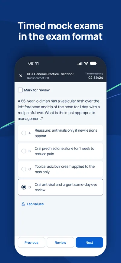
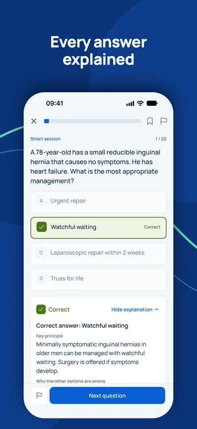
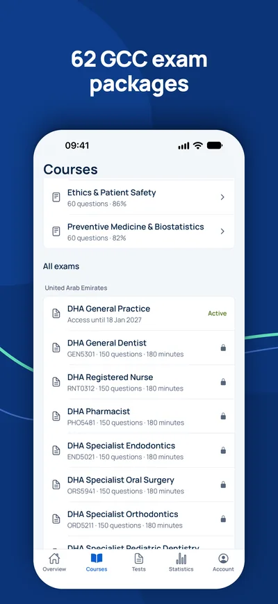
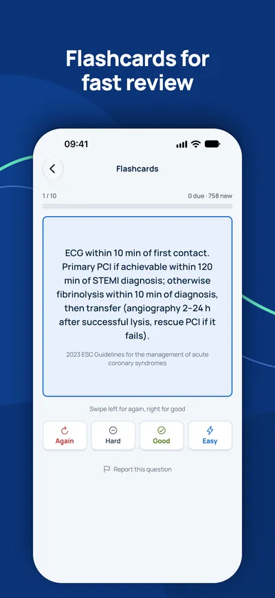
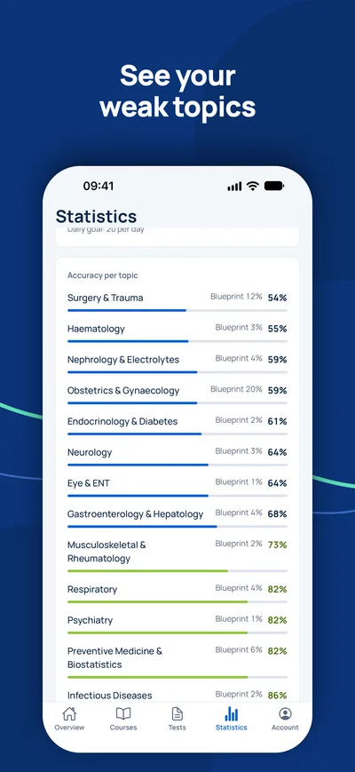
Is the exam your only open step?
Check first whether you need the exam at all and which title you can expect. A consultant answers your questions for free.
Eligibility check- 1Free consultation by video, phone or WhatsApp
- 2Eligibility check against the official requirements
- 3Your plan with fixed prices and clear next steps