
🇧🇭 NHRA
Fachzahnarzt Kieferorthopädie Bahrain
Fachzahnärztinnen und Fachzahnärzte in der Vorbereitung auf die Prüfungen in Kieferorthopädie bei DHA, DOH, Qatar DHP und SCFHS
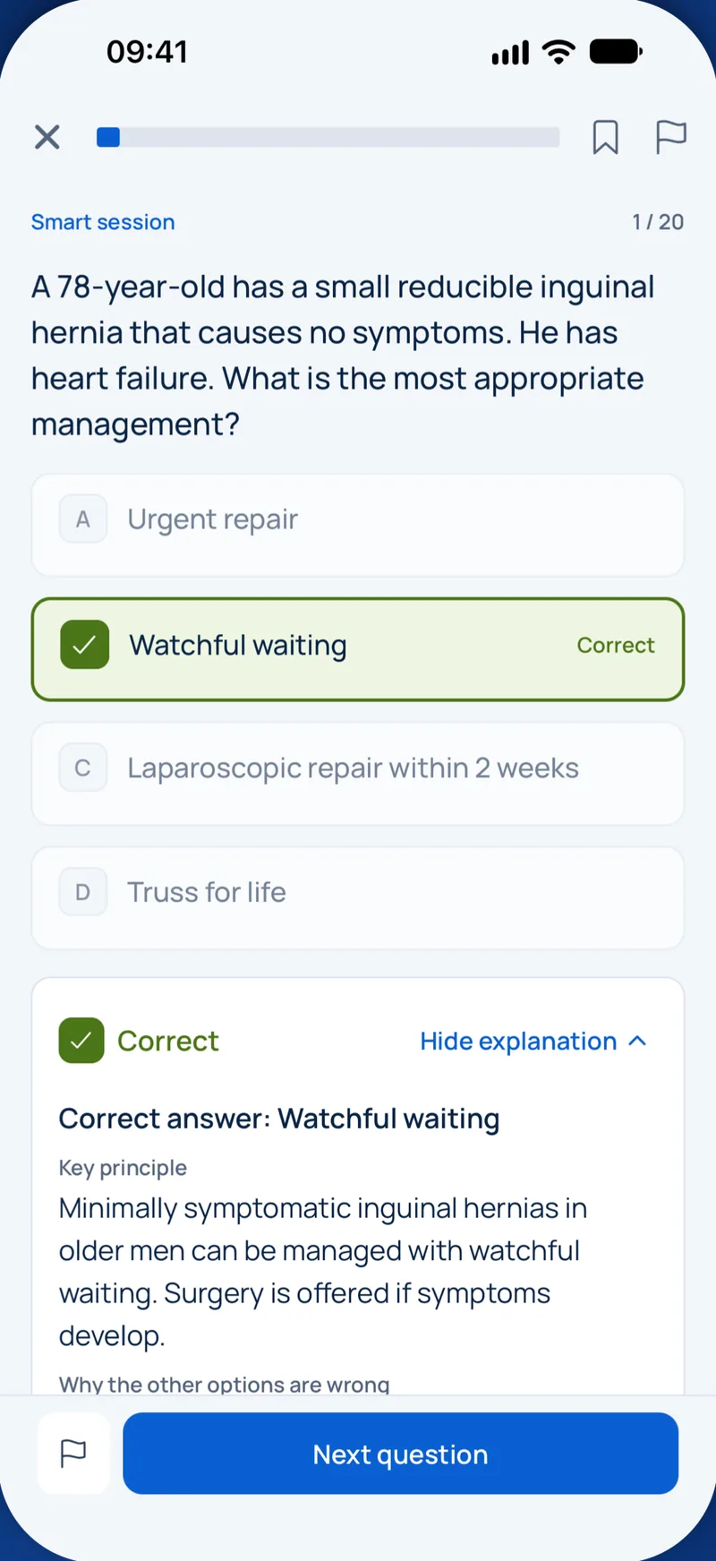

Beispielfrage
Which structure is the classic example of a secondary cartilage in the craniofacial skeleton?
Keys: 1–5 or A–E to choose, Enter to check and continue, F to mark.
Original MediFlow content, not official exam questions. Medical review in progress.
Syllabus and high-yield notes
1. Wachstum & Entwicklung · 15 % Anteil in der Prüfung · 5 lessons+
- Scammon-Kurven: Neurales Gewebe erreicht bis zum 6.–7. Lebensjahr etwa 90 % seines Wachstums, lymphatisches Gewebe übersteigt vor der Pubertät die Erwachsenengröße, allgemeines/skelettales und genitales Gewebe folgen einer pubertären S-Kurve. Im Gesicht endet zuerst das Breiten-, dann das Längen- (Tiefen-) und zuletzt das Höhenwachstum.
- Der Kondylenknorpel ist ein sekundärer Knorpel: Er wächst adaptiv unter funktionellen und Umwelteinflüssen, anders als primäre Knorpel (Epiphysenfugen, Synchondrosis sphenooccipitalis) mit eigenem Wachstumspotenzial.
- Die Maxilla wird durch Wachstum an den zirkummaxillären Suturen und den Schub der Schädelbasis nach unten und vorne verlagert, während ihre Vorderfläche resorptiv umgebaut wird; vertikales Alveolarfortsatzwachstum und Umbau des Gaumens ergänzen die Höhe.
- Björks Implantatstudien zeigten, dass die meisten Unterkiefer im Wachstum nach vorne (gegen den Uhrzeigersinn) rotieren; ein Großteil wird durch Umbau am Unterrand maskiert. Rückwärtsrotierer entwickeln eine große vordere Gesichtshöhe und Neigung zum offenen Biss.
- Die skelettale Reife lässt sich besser über Handröntgen (z. B. Sesambein, MP3-Stadien) oder die Halswirbelreifung (CVM, Baccetti CS1–CS6) als über das chronologische Alter bestimmen; der pubertäre Gipfel des Unterkieferwachstums liegt zwischen CS3 und CS4.
- Mädchen erreichen den pubertären Wachstumsschub etwa zwei Jahre früher als Jungen (rund 11–12 vs. 13–14 Jahre). Das vertikale Gesichtswachstum, besonders des Unterkiefers bei Männern, setzt sich bis in die späten Teenagerjahre und langsam ins Erwachsenenalter fort.
- Leeway-Space: Die Summe aus Milcheckzahn und Milchmolaren übertrifft die der Nachfolger um etwa 1,5 mm je Seite im Oberkiefer und 2,5 mm je Seite im Unterkiefer; er erlaubt die Mesialwanderung der Molaren von gerader Abschlusslinie zur Klasse I.
- Die Interkaninenbreite nimmt vor allem mit dem Durchbruch der bleibenden Schneide- und Eckzähne zu und steigt im Unterkiefer nach etwa 12–13 Jahren kaum noch; die Bogenlänge nimmt im Erwachsenenalter leicht ab und trägt zum späten unteren Frontengstand bei.
- Principles of craniofacial growth: patterns, cartilages and theories – Orthodontists treat growing patients, so they must know how much growth is left, where it happens and in which direction. This chapter covers the basic rules first: how different tissues grow at different times, how bone actually gets bigger, and the main theories that try to explain what controls facial growth. Later chapters apply these rules to the maxilla, the mandible, timing of treatment and the dentition.
- Growth of the cranial base, maxilla and mandible, and growth rotations – Once you know the basic mechanisms, the next step is to apply them to each part of the craniofacial skeleton. The cranial base is the platform on which the face hangs, the maxilla grows mainly by sutural growth and remodelling, and the mandible grows mainly at the condyle and ramus. The relation between vertical growth of the face and growth of the condyle decides whether the mandible rotates forward or backward, which in turn shapes the face.
- Timing growth: skeletal maturity, the pubertal spurt and adult growth – Many orthodontic decisions depend on timing. A functional appliance for mandibular retrusion works best during the pubertal spurt. Maxillary expansion is most skeletal when the patient is young. Orthognathic surgery for mandibular prognathism should wait until growth has stopped. Chronological age is a poor guide because children of the same age can be years apart in maturity. Clinicians therefore estimate skeletal age from radiographs and from other biological signs.
- Development of the dentition and dental arches – The dentition develops together with the jaws. Many features that look abnormal in a child, such as a midline gap or a flush molar relationship, are normal stages that resolve on their own. A specialist must know which features are transient and which need treatment. This chapter follows the dentition from the primary stage through the mixed dentition into the adult arch.
- Craniofacial syndromes, clefts and disturbances of growth – Most patients an orthodontist sees have a malocclusion that sits within normal variation. A small but important group have a recognisable disturbance of craniofacial growth: a syndrome, a sequence, a cleft, a joint disease or an injury that changed how the face grew. These patients need the orthodontist to understand which tissue failed, how that failure will play out over the remaining years of growth, and which other specialists must be involved. This chapter gives the growth logic behind the common conditions; the treatment chapter covers the clinical pathway for cleft care in more detail.
2. Diagnostik · 20 % Anteil in der Prüfung · 5 lessons+
- Eine problemorientierte Diagnose ordnet Befunde nach Dimension (sagittal, vertikal, transversal), Ebene (skelettal, dentoalveolär, Weichgewebe) sowie Ausrichtung/Symmetrie und priorisiert die Probleme vor der Therapiewahl.
- Die Wits-Analyse misst den Abstand der Lote von A- und B-Punkt auf die funktionelle Okklusionsebene; Mittelwert etwa 0 mm bei Frauen und −1 mm bei Männern. Sie umgeht die Nasion-Abhängigkeit von ANB, hängt aber von der Okklusionsebene ab.
- Der IOTN besteht aus einer Dental Health Component (Grade 1–5, schlimmstes Einzelmerkmal nach MOCDO: fehlende Zähne, Overjet, Kreuzbiss, Kontaktpunktverschiebung, Overbite) und einer Aesthetic Component (10 Fotos). DHC 4–5 bedeutet großen Behandlungsbedarf.
- Der PAR-Index misst die Abweichung von der Normokklusion; eine Reduktion um mindestens 30 % gilt als Verbesserung, eine Reduktion um 22 Punkte oder mehr als „stark verbessert“.
- DVT ist kein Routinebefund in der KFO. Es wird eingesetzt, wenn konventionelle Bildgebung eine konkrete Frage nicht beantwortet, z. B. verlagerter Eckzahn mit Verdacht auf Wurzelresorption, Spalt-Alveolarknochen oder komplexe skelettale Asymmetrie, mit kleinstmöglichem Volumen (ALARA/ALADAIP).
- Bolton-Analyse: anteriore Ratio etwa 77,2 %, Gesamtratio etwa 91,3 %. Eine Diskrepanz über etwa 1,5 mm ist klinisch relevant und beeinflusst das Finishing (Overjet, Overbite, Lücken).
- Ein funktioneller Zwangsbiss (Differenz zwischen habitueller und zentrischer Okklusion) muss vor der Diagnose erkannt werden: Pseudo-Klasse-III oder einseitiger Kreuzbiss mit Verschiebung sind oft dental bedingt, eine echte Asymmetrie bleibt in Zentrik bestehen.
- Die Weichgewebsdiagnostik lenkt die Planung: Nasolabialwinkel (etwa 90–110°), Lippenposition zur E-Linie nach Ricketts (oben −4, unten −2 mm beim Erwachsenen), Inzisivenexposition in Ruhe (etwa 2–4 mm) und Lachlinie. Extraktionsentscheidungen müssen das Profil berücksichtigen.
- The problem-oriented approach, clinical examination and records – Orthodontic diagnosis is not the same as naming an Angle class. Angle's classification describes only the sagittal relationship of the first molars. It says nothing about the skeleton behind the teeth, the vertical proportions of the face, the width of the arches, the soft tissues or the patient's concerns. Modern diagnosis is therefore problem-oriented: the clinician collects a database, turns it into a list of problems, ranks those problems, and only then thinks about treatment options. Treatment is chosen to solve the prioritised problems, not to normalise every number.
- Cephalometric analysis: skeletal and dental relationships – A lateral cephalogram is a standardised lateral skull radiograph taken with the head fixed in a cephalostat, the teeth in occlusion and the Frankfort plane horizontal. Its value is that the same landmarks can be identified on every film, allowing measurements to be compared with population norms and with the same patient over time. Cephalometric numbers are aids to diagnosis, not the diagnosis itself. Every value must be interpreted against the clinical picture, and a single abnormal number should never drive treatment.
- Space analysis, tooth size and characteristic malocclusion patterns – Space analysis compares the space available in an arch with the space required for the teeth. The space available is the arch perimeter from the mesial of one first molar to the mesial of the other, measured along the line of the ideal arch. The space required is the sum of the mesiodistal widths of the teeth in front of the first molars. A negative result means crowding, a positive result means spacing. Crowding is usually graded as mild (up to about 4 mm per arch), moderate (about 4 to 8 mm) and severe (more than about 8 mm). These bands are guides; the decision to extract also depends on incisor position, the profile, the curve of Spee and anchorage.
- Treatment need and outcome indices, and putting the diagnosis together – Indices turn clinical findings into numbers that can be compared between patients, clinicians and health systems. Two are essential for examinations: the Index of Orthodontic Treatment Need (IOTN), which ranks how much a patient needs treatment, and the Peer Assessment Rating (PAR), which measures how much a malocclusion deviates from normal and is used to judge treatment outcome. They answer different questions and must not be confused.
- Aetiology of malocclusion: genes, environment and local factors – Before choosing a treatment, it helps to ask why the malocclusion developed. The answer guides prognosis: a problem driven by a persistent habit or a soft tissue pattern will relapse if the cause continues, while one caused by a single local event, such as early loss of a primary molar, can often be fully corrected. Very few malocclusions have one clear cause. Most result from a genetically determined skeletal and dental pattern modified by environmental influences during growth.
3. Biomechanik · 20 % Anteil in der Prüfung · 5 lessons+
- Eine Kraft durch das Widerstandszentrum bewirkt Translation; eine exzentrische Kraft erzeugt ein Moment (Kraft × Hebelarm) und Rotation. Das Verhältnis Moment zu Kraft am Bracket bestimmt die Bewegungsart: etwa 7:1 kontrollierte Kippung, 10:1 Translation, über 10:1 Wurzelbewegung (für einen typischen Schneidezahn).
- Das Widerstandszentrum eines einwurzeligen Zahnes liegt etwa ein Drittel bis 40 % der Wurzellänge apikal des Alveolarkamms; bei Knochenabbau wandert es nach apikal, sodass dieselbe Kraft parodontal geschwächte Zähne stärker kippt.
- Druck-Zug-Theorie: Auf der Druckseite führt leichte Kraft zu direkter Resorption, starke Kraft zu Hyalinisierung (sterile Nekrose) und unterminierender Resorption mit Verzögerungsphase. RANKL/OPG und Entzündungsmediatoren (PGE2, IL-1) steuern die Osteoklastogenese.
- Die Steifigkeit eines runden Drahtes ist proportional zur vierten Potenz des Durchmessers (doppelter Durchmesser = 16-fach steifer) und umgekehrt proportional zur dritten Potenz der Spannweite; für die Reichweite gilt das Umgekehrte. Bei Vierkantdraht steigt sie mit Breite × Höhe³ in Biegerichtung.
- Nickel-Titan zeigt Superelastizität (spannungsinduzierte martensitische Umwandlung) mit flacher Entlastungskurve sowie Formgedächtnis; thermisch aktivierte Drähte haben eine Austenit-Endtemperatur nahe der Mundtemperatur. TMA (Beta-Titan) hat etwa 40 % der Stahlsteifigkeit und ist bieg- und schweißbar.
- Statisch bestimmte Systeme (Ein-Kraftpaar: Cantilever, Intrusionsbogen mit Einpunktligatur) lassen Kräfte und Momente bekannt sein; Zwei-Kraftpaar-Systeme (Draht in zwei Brackets) sind statisch unbestimmt. Burstones Geometrieklassen beschreiben die Kraftsysteme eines geraden Drahtes zwischen zwei Brackets.
- Verankerung ist der Widerstand gegen unerwünschte Bewegung. Sie wird verstärkt durch mehr Zähne, differenzielle Momente, extraorale Kräfte oder skelettale Verankerung (Minischrauben, Miniplatten), die richtig gesetzt absolute Verankerung bietet; die Primärstabilität hängt von Kortikalisdicke und Eindrehmoment (etwa 5–10 Ncm) ab.
- Reibung beim Gleiten besteht aus klassischer Reibung, Binding (Angulation zweiter Ordnung über den kritischen Kontaktwinkel) und Notching; klinisch dominiert Binding. Selbstligierende Brackets senken die klassische Reibung bei dünnen Runddrähten, nicht aber das Binding.
- Forces, moments and the centre of resistance – Every orthodontic appliance does one thing: it delivers a force system to a tooth. Whether the tooth tips, moves bodily, rotates or moves its root depends not on the appliance type but on the forces and moments it receives relative to one special point, the centre of resistance. If you understand this chapter, most biomechanics questions become simple geometry.
- Biology of tooth movement and force magnitude – Mechanics decides where a tooth wants to go; biology decides whether and how fast it gets there. Orthodontic tooth movement is a remodelling process of the PDL and alveolar bone: bone is resorbed in the direction of movement and formed behind the tooth. Knowing how force magnitude changes this biology explains lag phases, pain, root resorption and the recommended force ranges.
- Wires, materials and friction – Archwires store energy when they are deflected and release it as force on the teeth. Choosing a wire means choosing how much force it delivers per millimetre of activation, how far it can be bent before deforming permanently, and how easy it is to shape. These are three related but different properties.
- Force systems in practice and anchorage – In the mouth, appliances connect teeth to other teeth, so every force system has an active part and a reactive part. Good biomechanics means predicting both, then using or neutralising the reaction. This chapter applies the principles of the first three chapters to real appliances and to anchorage.
- Adverse effects of tooth movement: root resorption, pulp, periodontium, enamel and drugs – Every orthodontic force that moves a tooth also stresses the root, the pulp, the periodontal ligament and the alveolar bone. Most of these effects are mild and reversible. Some are not. A specialist must be able to predict who is at risk, choose mechanics that reduce harm, recognise problems early and explain them to patients. This chapter looks at the tissue side of biomechanics.
4. Apparaturen · 15 % Anteil in der Prüfung · 5 lessons+
- Vorprogrammierte Edgewise-Systeme (Andrews Straight-Wire, Roth, MBT) enthalten Angulation, Torque und In-out im Bracket; MBT hat mehr palatinalen Wurzeltorque an den oberen Schneidezähnen (+17°) und weniger Torque an den unteren (−6°) als Roth, um die Gleitmechanik auszugleichen.
- Die Torque-Übertragung hängt vom Slot-Spiel ab: Ein 0,019 × 0,025-Zoll-Stahldraht in einem 0,022-Zoll-Slot hat etwa 10–12° Spiel, sodass der volle Torque nicht übertragen wird.
- Funktionskieferorthopädische Geräte (Twin Block, Herbst, Aktivator, Fränkel) korrigieren die Klasse II überwiegend dentoalveolär: Retrusion der oberen und Protrusion der unteren Schneidezähne, mit kleinem, variablem skelettalem Unterkiefereffekt (etwa 1–2 mm).
- Headgear: Zervikalzug extrudiert Molaren und kann den Biss öffnen (für Low-Angle-Fälle), High-Pull intrudiert Molaren und unterstützt die vertikale Kontrolle bei High-Angle-Fällen; kombinierter Zug liegt dazwischen. Sicherheitsvorrichtungen (Sicherheitsmodul, verriegelter Gesichtsbogen) sind Pflicht.
- Die forcierte Gaumennahterweiterung (Hyrax, Haas) öffnet die Sutura palatina mediana mit etwa 0,25–0,5 mm Aktivierung pro Tag; ein Mittendiastema entsteht. Minischraubengestützte Geräte (MARPE) erweitern die skelettale Dehnung auf späte Jugendliche und Erwachsene.
- Aligner bewegen Zähne in Schritten von etwa 0,25 mm pro Schiene mithilfe von Attachments; Extrusion, Rotation runder Zähne und körperliche Bewegungen sind weniger vorhersagbar. Nachkorrekturen sind häufig.
- Herausnehmbare Geräte: Adams-Klammer an ersten Molaren, Southend-Klammer an Schneidezähnen; Federn aus 0,5-mm-Stahl bewegen einzelne Zähne kippend. Körperliche Bewegungen oder größere Rotationen sind nicht möglich.
- Gesichtsmaske (Delaire) zur Protraktion wirkt am besten vor etwa 10 Jahren, ideal im frühen Wechselgebiss; Kräfte von etwa 300–500 g je Seite, 12–14 Stunden täglich, etwa 30° nach unten zur Okklusionsebene im Eckzahnbereich. Knochenverankerte Protraktion (Miniplatten mit Klasse-III-Gummizügen) wirkt auch bei älteren Kindern.
- Fixed appliances: brackets, prescriptions and torque – A fixed appliance is a set of brackets bonded to the teeth (and bands or tubes on the molars) connected by an archwire. The wire is elastically deformed when it is engaged into brackets on misaligned teeth. As it tries to return to its original shape, it delivers forces and moments to the teeth. The bracket is the interface that translates wire shape into tooth position, so understanding what is built into the bracket explains most of the mechanics questions examiners ask.
- Removable appliances, space maintenance, expansion and anchorage devices – A removable appliance consists of an acrylic baseplate, retentive components (clasps), active components (springs, screws, elastics) and anchorage from the baseplate and palate. Its great limitation is that it holds a tooth with a single point of force. A single force through the crown, away from the centre of resistance, produces tipping. To produce bodily movement or controlled root movement, a couple (two forces) is needed, and a removable appliance cannot apply one effectively. Therefore removable appliances move teeth by tipping only and cannot correct significant rotations, close extraction spaces bodily or control torque.
- Class II growth modification: functional appliances and headgear – Growth modification aims to change the relationship of the jaws while the patient is still growing. In Class II malocclusion, the typical goals are to restrain forward maxillary growth or redirect it, and to encourage the mandible into a more forward position. Timing is important. Most evidence shows that treatment during the adolescent growth spurt achieves the same result in one phase that early two-phase treatment achieves, with less total treatment time. Early treatment is justified mainly for children with a large overjet who are at risk of incisor trauma or who are teased.
- Class III protraction, miniscrews and clear aligners – Skeletal Class III can result from maxillary deficiency, mandibular excess or both. In many children the main component is a small or retrusive maxilla. The maxilla is attached to the cranial base through circummaxillary sutures, which remain responsive to orthopaedic force in young children. Protraction therefore works best early, before about 10 years and ideally in the early mixed dentition.
- Patient safety, ethics and professionalism in orthodontics – Orthodontic treatment is elective, usually performed on healthy young people, and lasts for years. That combination raises the bar for safety and ethics. The patient is often a minor, the benefit is partly aesthetic and psychological, and harm from the treatment itself, such as decalcification, root resorption or an eye injury from headgear, can be permanent. Licensing examinations in the Gulf now test this area explicitly, alongside the clinical content.
5. Behandlung · 20 % Anteil in der Prüfung · 5 lessons+
- Eine zweiphasige Klasse-II-Behandlung (früh funktionell, dann festsitzend) liefert kein besseres skelettales Endergebnis als eine einphasige Behandlung im Jugendalter, senkt aber bei großem Overjet das Risiko eines Frontzahntraumas.
- Palatinal verlagerte Eckzähne: Ist der Eckzahn mit 10–11 Jahren nicht vestibulär tastbar, wird geröntgt; die Extraktion des Milcheckzahns zwischen 10 und 13 Jahren verbessert die Chance auf spontanen Durchbruch, wenn die Krone weniger als die Hälfte der seitlichen Wurzel überlappt; GNE oder Headgear können ergänzen.
- Kamouflage versus Chirurgie: Bei Klasse II wird beim Erwachsenen meist ab einem Overjet über etwa 10 mm, starker skelettaler Diskrepanz oder inakzeptablem Profil operiert; bei Klasse III ist Kamouflage begrenzt, wenn die umgekehrte Stufe groß ist und die Schneidezähne bereits kompensiert sind.
- Die präoperative Kieferorthopädie dekompensiert die Schneidezähne, nivelliert und koordiniert die Bögen; Profil und Overjet verschlechtern sich vorübergehend. Surgery-first-Protokolle nutzen postoperativ das regionale Akzelerationsphänomen.
- Offener Biss beim Erwachsenen mit langem Gesicht: Optionen sind Molarenintrusion mit skelettaler Verankerung, Extrusion der Schneidezähne nur bei geringer Inzisivenexposition oder Le-Fort-I-Impaktion. Zungenlage und Habits müssen für die Stabilität berücksichtigt werden.
- Hypodontie (am häufigsten untere zweite Prämolaren, dann obere seitliche Schneidezähne): Lückenschluss (Eckzahnsubstitution) oder Lückenöffnung für Implantate nach Profil, Eckzahnform/-farbe, Seitenzahnokklusion und Engstand entscheiden; Implantate erst nach Wachstumsabschluss.
- Interdisziplinäre Erwachsenen-KFO setzt zunächst parodontale Stabilität voraus (keine aktiven Taschen), leichte Kräfte und häufig festsitzende Retention; nach parodontaler Regeneration wird meist 3–6 Monate gewartet, bevor Zähne in Defekte bewegt werden.
- Die Versorgung von Lippen-Kiefer-Gaumenspalten erfolgt im Team: Die sekundäre Osteoplastik erfolgt idealerweise vor dem Durchbruch des bleibenden Eckzahns (etwa 8–11 Jahre, Eckzahnwurzel zu einer Hälfte bis zwei Dritteln ausgebildet); die Oberkiefervorverlagerung nach Wachstumsabschluss.
- Interceptive and early treatment in the mixed dentition – Interceptive orthodontics means treatment in the primary or mixed dentition that removes a cause of malocclusion or stops a developing problem from getting worse. It is not the same as early comprehensive treatment. The key question for every early intervention is simple: does treating now give a benefit that cannot be obtained later, at lower cost and burden? Examiners test whether you know which early treatments have evidence of benefit and which do not.
- Impacted canines and hypodontia – The maxillary permanent canine has the longest path of eruption of any tooth and erupts late, at about 11 to 12 years. It is therefore often displaced, most commonly palatally (around 85 percent of displaced upper canines in European populations). Palatal displacement is linked with small, peg-shaped or missing lateral incisors and has a familial pattern, which supports the guidance theory: the lateral incisor root guides the canine down. Buccal displacement is more often due to crowding.
- Camouflage, orthognathic surgery and distraction – When a malocclusion has a significant skeletal cause after growth has stopped, there are two broad choices. Orthodontic camouflage moves the teeth to hide the skeletal discrepancy: the jaws stay where they are, and the incisors are tipped to compensate. Orthognathic surgery corrects the jaw positions, combined with orthodontics to place the teeth correctly over the new skeletal base. Growth modification, the third option, is only possible in growing patients.
- Adult interdisciplinary care and cleft lip and palate – Adults now form a large share of orthodontic patients. Compared with adolescents they have no growth to help, more restorations and missing teeth, slower tissue response and more often periodontal disease. Adult treatment is therefore often interdisciplinary: orthodontics prepares the mouth for periodontal, restorative or implant work, or is limited to specific goals.
- Special considerations: medical conditions, TMD, sleep apnoea and pregnancy – Orthodontic patients are not always healthy teenagers. Adults and children with medical conditions, temporomandibular disorders or sleep-disordered breathing increasingly ask for treatment. The orthodontist must decide whether treatment is safe, how it must be modified and which other clinicians need to be involved. The guiding question is simple: does the condition change the risk of treatment, the response of the tissues, or the patient's ability to cope with appliances?
6. Retention & Stabilität · 10 % Anteil in der Prüfung · 5 lessons+
- Rezidiv ist die Tendenz der Zähne zur Rückkehr in die Ausgangslage; Ursachen sind Rückstellkräfte von PDL und gingivalen Fasern (suprakrestale Fasern bauen sich langsam um, etwa ein Jahr oder länger), Weichgewebsdruck und Restwachstum. Langzeit- bzw. unbefristete Retention ist heute Standard.
- Littles Langzeitstudien: Etwa zwei Drittel der Extraktionsfälle zeigten 10 Jahre nach Retentionsende eine unbefriedigende Ausrichtung der unteren Schneidezähne; das Rezidiv war aus den Ausgangsdaten nicht vorhersagbar.
- Geklebte Retainer sichern die Ausrichtung gut, versagen aber (Ablösung, Drahtbruch) in etwa einem Drittel der Fälle über mehrere Jahre; mit flexiblen Mehrstrangdrähten kann unerwünschter Torque („X-“ oder Twist-Effekt) auftreten.
- Tiefgezogene (Essix-) und Hawley-Retainer bieten bei nächtlichem Tragen ähnliche Stabilität; Essix-Retainer werden ästhetisch besser akzeptiert, verschleißen aber schneller.
- Rotationen rezidivieren stark; die zirkumferente suprakrestale Fibrotomie (CSF) mindert das Rotationsrezidiv vor allem im Oberkiefer und erfolgt gegen Behandlungsende.
- Die untere Interkaninenbreite ist nur stabil, wenn sie nicht erweitert wurde; eine Dehnung im unteren Eckzahnbereich rezidiviert meist.
- Die Korrektur eines offenen Bisses rezidiviert bei einem relevanten Anteil (etwa 20–35 %), besonders bei Zungenfehlfunktion; die Tiefbisskorrektur teilweise; die Klasse-III-Korrektur rezidiviert durch spätes Unterkieferwachstum.
- Patienten müssen vor Behandlungsbeginn erfahren, dass Retention eine lebenslange Aufgabe ist, Kontrollen nötig sind und defekte Retainer schnell repariert werden müssen.
- Why teeth relapse: the biology of stability and the case for long-term retention – Retention is the phase of orthodontic care that holds the teeth in their corrected positions after active tooth movement ends. Relapse is the tendency of teeth to return towards their original positions. Retention is not an optional extra at the end of treatment. It is part of the treatment plan from the first consultation, because without it a large share of corrections will partly undo themselves.
- Retainers: types, evidence, choice and maintenance – A retainer is any appliance that holds teeth after active treatment. There are two broad groups. Removable retainers are taken out by the patient and depend on cooperation. Fixed (bonded) retainers are glued to the lingual or palatal surfaces of the teeth and work without cooperation, but they need maintenance and good hygiene. No single retainer is best for every patient. The choice depends on the original malocclusion, the movements performed, the periodontal condition, oral hygiene and the patient's wishes.
- Retention tailored to the problem: rotations, spaces, vertical and sagittal corrections, adults and consent – Good retention planning starts by asking which movements are most likely to relapse in this patient. The retainer and any adjunct are then chosen to resist those specific forces. This chapter goes through the common situations that examiners use.
- Retention in special situations: expansion, implants, restorative work, clefts and surgery – The general principles of retention apply to every patient: teeth tend to return toward their original positions, soft tissues and continuing growth push them, and the safest plan is long-term retention. Some situations add specific risks or require coordination with other clinicians. This chapter covers the most common ones and explains how the retention plan should be adapted.
- Retainer failure, relapse management, retreatment and responsibility – Even with a good plan, retainers break, get lost or are not worn, and some teeth move. A specialist needs a systematic approach to these events: identify what has moved and why, decide whether intervention is needed, discuss options with the patient honestly, and prevent the same failure from happening again. This chapter also covers who is responsible for retention over the long term.
Wachstum & Entwicklung
Lesson 1Principles of craniofacial growth: patterns, cartilages and theories
Orthodontists treat growing patients, so they must know how much growth is left, where it happens and in which direction. This chapter covers the basic rules first: how different tissues grow at different times, how bone actually gets bigger, and the main theories that try to explain what controls facial growth. Later chapters apply these rules to the maxilla, the mandible, timing of treatment and the dentition.
Two words are used constantly. Growth means an increase in size or number. Development means increasing complexity and maturity. A child can grow taller without much development, and a jaw can mature without getting much bigger. Exams often test growth pattern (the proportion and direction of growth over time) rather than absolute size.
Scammon's growth curves (1930) show that the body does not grow as one unit. Each tissue type follows its own curve, expressed as a percentage of adult size reached at each age. The curves explain why the skull is large in a baby and the face is small, and why the face catches up during adolescence.
| Tissue type (Scammon) | Curve shape | Practical meaning |
|---|---|---|
| Neural (brain, cranial vault, eyes) | Very early; about 90 % of adult size by age 6–7 | The cranial vault and anterior cranial base are almost finished in early childhood |
| Lymphoid (tonsils, adenoids, thymus) | Rises to about 200 % of adult size before puberty, then shrinks | Large adenoids in children are normal; they regress in adolescence |
| General / somatic (skeleton, muscles, jaws) | S-shaped: fast in infancy, slow in childhood, pubertal spurt, then stop | The jaws, especially the mandible, follow this curve and show a pubertal spurt |
| Genital | Almost flat until puberty, then steep rise | Marks sexual maturation; links to the timing of the facial spurt |
The face lies between the neural and general curves. The upper face, attached to the cranium, finishes earlier. The mandible, furthest from the brain, behaves most like the general skeleton and has the largest and latest pubertal spurt. This is the cephalocaudal gradient of growth: structures nearer the head mature earlier, those further away later.
There is also a dimensional gradient in the face. Width (transverse) is completed first, then length or depth (anteroposterior), and height (vertical) last. Intercanine and maxillary width are largely fixed before the adolescent spurt, while vertical growth of the face and alveolar processes continues into the late teens and, slowly, into adulthood. This order has direct clinical consequences: transverse problems should be corrected early, and vertical problems may relapse late.
How does bone grow? Bone cannot grow by expanding from inside (interstitial growth) because its matrix is mineralised. It grows only by adding bone on a surface (apposition) and removing it elsewhere (resorption). Cartilage, in contrast, can grow interstitially. Facial bones form either within membrane (intramembranous: cranial vault, maxilla, most of the mandible) or by replacing a cartilage model (endochondral: cranial base, limbs).
Three kinds of growth site matter in the face: sutures (fibrous joints between bones, such as the circummaxillary sutures), cartilages (cranial base synchondroses, nasal septum, mandibular condyle) and the periosteal and endosteal surfaces, where remodelling occurs.
A key distinction is between primary and secondary cartilage. Primary cartilage comes from the original cartilaginous skeleton of the embryo. Its chondrocytes divide in organised columns and it has its own intrinsic growth potential; when transplanted it keeps growing. Examples are the epiphyseal plates of long bones, the spheno-occipital synchondrosis and the nasal septal cartilage. Secondary cartilage develops later, from a periosteal-like layer, and grows adaptively in response to local function and mechanical environment. The mandibular condylar cartilage is the classic secondary cartilage. Its prechondroblasts lie in a fibrous covering, it grows by apposition rather than ordered interstitial columns, and it responds to mandibular posture and function. That is the biological reason why functional appliances were thought to be able to influence it, although the clinical effect on final mandibular length is modest.
| Feature | Primary cartilage | Secondary cartilage (condyle) |
|---|---|---|
| Origin | Embryonic cartilage skeleton | Arises secondarily from periosteal-like tissue |
| Growth control | Intrinsic, genetically driven | Adaptive, responds to function and environment |
| Cell arrangement | Columns, interstitial growth | Undifferentiated cells in a fibrous covering, appositional |
| Examples | Epiphyseal plates, spheno-occipital synchondrosis, nasal septum | Mandibular condyle |
The spheno-occipital synchondrosis is the most important growth site of the cranial base after early childhood. Growth here lengthens the posterior cranial base and carries the maxilla forward. It closes in late adolescence, roughly 13–15 years in girls and 15–17 years in boys. The other synchondroses close much earlier.
Enlow described two separate processes that together move a bone. Remodelling (cortical drift) is apposition on one surface and resorption on another, which changes the shape and size of a bone and moves its surfaces. Displacement (translation) is the movement of the whole bone away from its neighbours while it grows. Primary displacement is caused by the bone's own growth, for example the maxilla pushed forward as bone is added at its posterior sutures. Secondary displacement is caused by growth of a neighbouring structure, for example the maxilla carried forward by lengthening of the anterior cranial base.
Enlow's V principle describes how many facial bones grow. Picture a V-shaped region, such as the palate or the coronoid process. Bone is deposited on the inner surface and resorbed on the outer surface. The V therefore moves toward its wide end and enlarges at the same time.
Theories of growth control try to answer one question: what is the primary determinant? The genetic (bone) theory, associated with Sicher, saw all skeletal tissues, including sutures, as primary growth centres. This was disproved: sutures react to tension and do not drive growth themselves. Scott's cartilage theory gave the nasal septum and other cartilages the leading role; septal cartilage does appear to pace midface growth, and septal damage in animals reduces midface growth. Moss's functional matrix hypothesis states that bone growth is secondary. The soft tissues and spaces (functional matrices) grow and function, and the bones respond. Periosteal matrices, like muscles, act directly on bone by remodelling; capsular matrices, like the brain and the oronasopharyngeal space, move bones passively by displacement. Van Limborgh combined these ideas: intrinsic genetic factors, local epigenetic factors such as cartilage and muscle, and general environmental factors all play a part. Most clinicians use this multifactorial view.
Sources: Proffit WR, Fields HW, Larson BE, Sarver DM. Contemporary Orthodontics, 6th ed. Elsevier, 2019 (chapters 2 and 4). · Enlow DH, Hans MG. Essentials of Facial Growth, 2nd ed. Needham Press, 2008. · Moss ML. The functional matrix hypothesis revisited. Am J Orthod Dentofacial Orthop 1997;112:8–11, 221–226, 338–342, 410–417. · Scammon RE. The measurement of the body in childhood. In: Harris JA et al. The Measurement of Man. University of Minnesota Press, 1930.
Zur NHRA Fachzahnarzt Kieferorthopädie-Prüfung
Wie viele Fragen hat die NHRA Fachzahnarzt Kieferorthopädie-Prüfung?+
nicht veröffentlicht. Genaue Fragenzahl, Zeit und Bestehensgrenze stehen in Ihrer Buchung.
Welche Themen haben das größte Gewicht?+
Die größten Themen sind Diagnostik (20 %), Biomechanik (20 %), Behandlung (20 %). Der Kurs umfasst 6 Themen mit insgesamt 30 Kapiteln.
Wie bereite ich mich auf die NHRA Fachzahnarzt Kieferorthopädie-Prüfung vor?+
Arbeiten Sie die Kapitel Thema für Thema durch, wiederholen Sie die Lernnotizen und üben Sie dann mit zeitgesteuerten Probeprüfungen im Format Ihrer Behörde.
Fragen
Wie lange habe ich Zugang?+
Der Zugang gilt 90 Tage ab Zahlung für das gewählte Land und den gewählten Beruf.
Welche Geräte kann ich nutzen?+
Jeden Browser auf Computer, Tablet oder Handy sowie die MediFlow-App mit demselben Konto.
Kann ich das Geld zurückbekommen?+
Bitte lesen Sie vor dem Kauf unsere Erstattungsrichtlinie. Erstattungsrichtlinie

iPhone · iPad
GCC Medical Exam Prep
Bereiten Sie sich auf Ihrem iPhone auf DHA, DOH, MOHAP, SCFHS und weitere Zulassungsprüfungen am Golf vor.

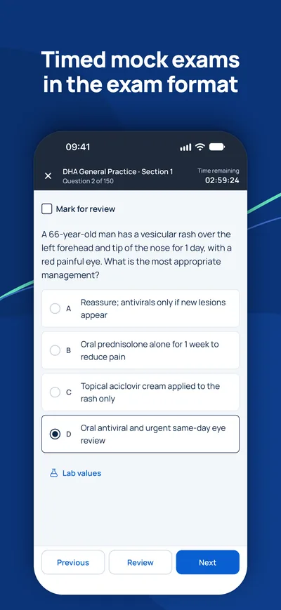
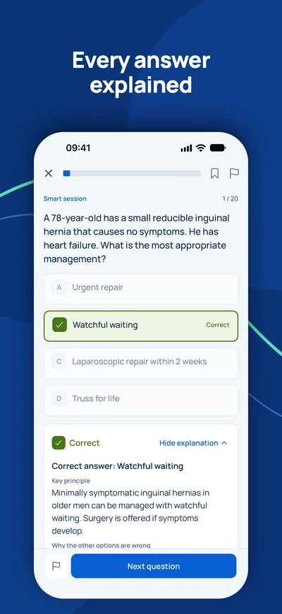
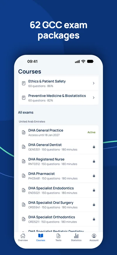


Ist die Prüfung Ihr einziger offener Schritt?
Prüfen Sie zuerst, ob Sie die Prüfung überhaupt brauchen und welchen Titel Sie erwarten können. Ein Berater beantwortet Ihre Fragen kostenlos.
Eignungs-Check- 1Kostenlose Beratung per Video, Telefon oder WhatsApp
- 2Eignungs-Check gegen die offiziellen Anforderungen
- 3Ihr Plan mit Festpreisen und klaren nächsten Schritten