
🇴🇲 OMSB
Fachzahnarzt Mund-, Kiefer- und Gesichtschirurgie Oman
Fachzahnärztinnen und Fachzahnärzte in der Vorbereitung auf die Prüfungen in Mund-, Kiefer- und Gesichtschirurgie bei DHA, DOH, Qatar DHP und SCFHS
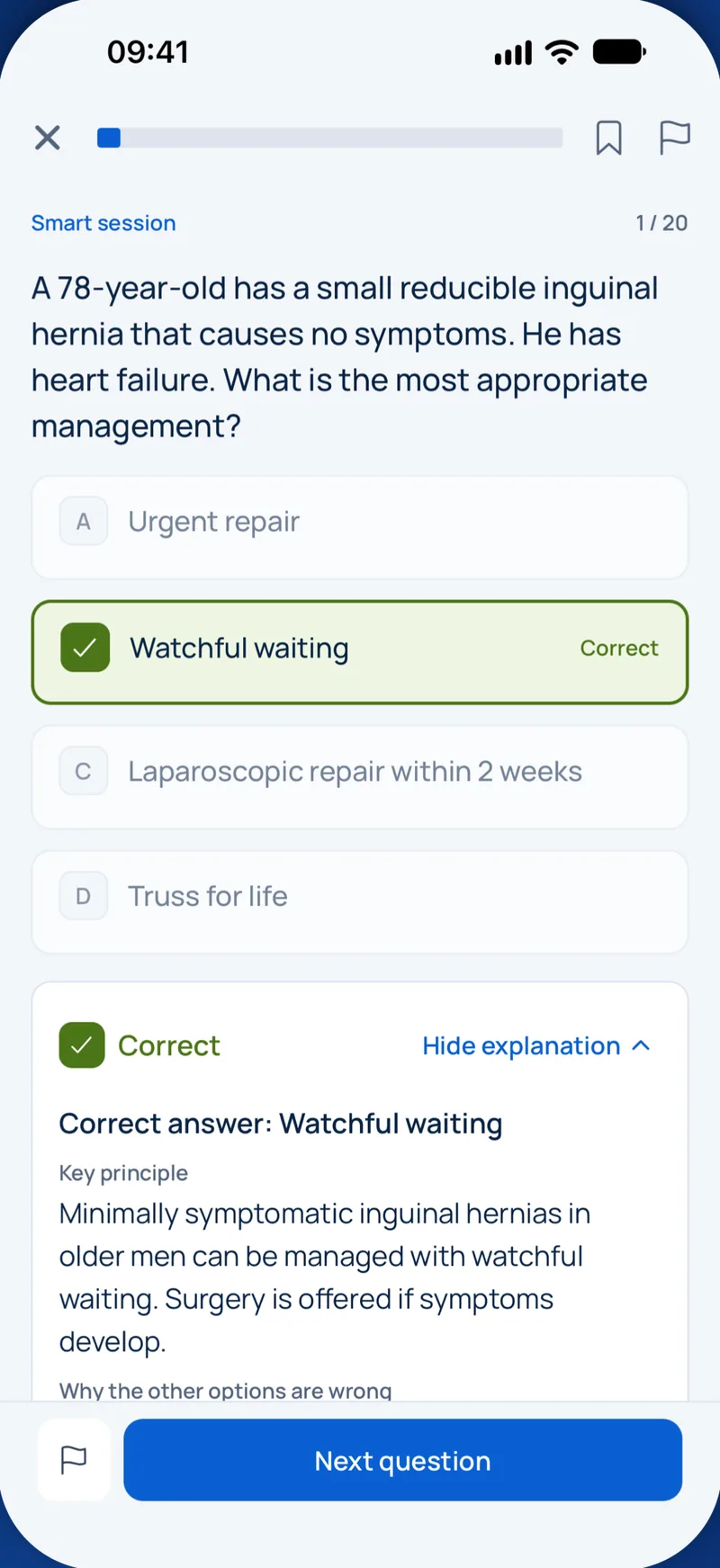
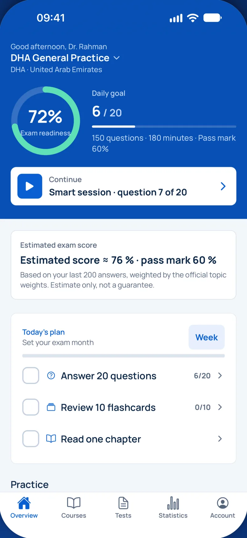
Beispielfrage
Which radiographic sign on a panoramic radiograph is most strongly associated with direct contact between a mandibular third molar root and the inferior alveolar canal?
Keys: 1–5 or A–E to choose, Enter to check and continue, F to mark.
Original MediFlow content, not official exam questions. Medical review in progress.
Syllabus and high-yield notes
1. Extraktion & Retention · 15 % Anteil in der Prüfung · 5 lessons+
- NICE TA1 lehnt die prophylaktische Entfernung symptomfreier Weisheitszähne ab; Indikationen sind u. a. nicht versorgbare Karies, Pulpa- oder periapikale Pathologie, Abszess, Osteomyelitis, Resorption, Fraktur, Zyste oder Tumor und eine zweite (oder eine schwere) Perikoronitis.
- Rood-Zeichen der engen Wurzel-Kanal-Beziehung: Aufhellung, Ablenkung und Verschmälerung der Wurzel, dunkle bifide Spitze; am Kanal Unterbrechung der Kortikalislinie, Umlenkung und Einengung. Umlenkung, Aufhellung der Wurzel und Unterbrechung der weißen Linie sind am riskantesten.
- Ein DVT ist indiziert, wenn das OPG ein Hochrisikozeichen zeigt, um Operation oder Koronektomie zu planen; es ist keine Routineuntersuchung vor jeder Extraktion.
- Winter beschreibt die Achsneigung (mesioangulär, vertikal, distoangulär, horizontal); im Unterkiefer ist mesioangulär am häufigsten und am leichtesten, distoangulär am schwersten. Im Oberkiefer ist distoangulär am leichtesten.
- Die meisten Lingualisschäden nach Weisheitszahnoperation erholen sich in Monaten; eine vollständige Anästhesie ohne Besserung über etwa 3 Monate ist die übliche Indikation zur mikrochirurgischen Revision.
- Eine Mund-Antrum-Verbindung unter etwa 2 mm heilt meist unter Koagel und Schonung; größere Defekte werden primär verschlossen (Rehrmann-Plastik, Bichat-Fettpfropf oder palatinaler Rotationslappen).
- Ein noch am Periost haftendes Tuberfragment wird reponiert und geschient; der Zahn wird 6–8 Wochen später chirurgisch entfernt.
- Bei der Koronektomie bleiben vitale Wurzeln mindestens 3 mm unter dem Knochenrand; Wurzelwanderung ist zu erwarten, eine Nachoperation selten nötig. Kontraindikationen sind mobile oder infizierte Wurzeln.
- Principles of exodontia: assessment, instruments and technique – Exodontia is the removal of teeth. At specialist level the examiner is not interested in whether you can use forceps; they want to know whether you can predict difficulty, choose the right technique before you start, and recognise when a simple extraction should become a surgical one. Most complications are decided in the planning phase, not in the chair.
- Impacted third molars: indications, classification, imaging and surgery – An impacted tooth is one that cannot erupt into a functional position because of bone, soft tissue or another tooth in its path. Lower third molars are the most commonly impacted teeth. The key specialist skills are deciding whether the tooth should come out at all, describing it in standard terms, judging the risk to the inferior alveolar nerve, and choosing the operation that minimises harm.
- Complications of exodontia: nerves, sinus, displaced teeth and dry socket – Complications are the core of specialist exams because they test judgement. For each one you should know how to prevent it, how to recognise it, what to do on the day, and when to refer.
- Local anaesthesia and pain management in oral surgery – Almost every oral surgical procedure starts with local anaesthesia, and many problems that candidates are asked about (failed blocks, toxicity, nerve injury, postoperative pain) come back to how well the anaesthetic and analgesic plan was built. This chapter explains how local anaesthetics work, how to choose the agent and dose, how the main blocks are given, why they fail, and how to control pain after surgery.
- Preprosthetic, implant and bone augmentation surgery – After teeth are lost, the alveolar ridge resorbs. The greatest loss happens in the first six to twelve months, and horizontal width is lost faster than height. Preprosthetic surgery prepares the mouth for a denture or implants: it removes obstacles, deepens the sulcus and, today mostly, rebuilds bone so that implants can be placed in the right prosthetic position. This chapter covers soft and hard tissue preprosthetic procedures, socket preservation, the basics of implant surgery, and the main augmentation techniques with their complications.
2. Infektionen · 15 % Anteil in der Prüfung · 5 lessons+
- Odontogene Infektionen sind polymikrobiell (vergrünende Streptokokken, Prevotella, Fusobacterium, Peptostreptococcus). Herdsanierung durch Drainage und Beseitigung der Ursache ist die Hauptbehandlung; Antibiotika ergänzen.
- Nach Flynn: geringes Risiko (Vestibulum, bukkal, Fossa canina, infraorbital), mittleres (submandibulär, submental, sublingual, pterygomandibulär, submasseterisch, temporal), hohes Risiko (parapharyngeal, retropharyngeal, prätracheal, Mediastinum, Danger Space).
- Die Ludwig-Angina ist eine beidseitige Phlegmone der submandibulären, sublingualen und submentalen Logen; die Atemwegssicherung (wache fiberoptische Intubation oder Tracheotomie in Lokalanästhesie) geht der Inzision voraus.
- Amoxicillin bzw. Penicillin V ist Mittel der Wahl; bei sich ausbreitender Infektion plus Metronidazol; Clindamycin bei Penicillinallergie, mit C.-difficile-Risiko und zunehmender Resistenz.
- Die Sinus-cavernosus-Thrombose aus Oberkieferfrontzähnen breitet sich über klappenlose V. facialis, angularis und ophthalmica oder über den Plexus pterygoideus aus; Zeichen: Protrusio, Chemosis, Ophthalmoplegie (zuerst N. VI) und Übergreifen auf das andere Auge.
- Die absteigende nekrotisierende Mediastinitis wird mit Kontrastmittel-CT von Hals und Thorax gesichert; sie erfordert dringliche zervikale und thorakale Drainage und Breitspektrumantibiotika.
- Die chronisch eitrige Osteomyelitis des Unterkiefers erfordert Sequestrotomie, ggf. Dekortikation und langfristige kulturgerechte Antibiose; die Garré-Osteomyelitis bei Kindern zeigt zwiebelschalenartigen Periostknochen und heilt nach Behandlung der Zahnursache.
- Die nekrotisierende Fasziitis des Halses zeigt Krepitation, livide Haut, unverhältnismäßige Schmerzen und Allgemeintoxizität; Therapie ist notfallmäßiges radikales Débridement plus Breitspektrumantibiose.
- Odontogenic infection: microbiology, stages and fascial spaces – Odontogenic infections start from a tooth or its supporting tissues, most often a necrotic pulp, a periodontal pocket or pericoronitis. Pus follows the path of least resistance. It perforates the thinnest cortical plate near the root apex and then enters whichever soft-tissue compartment lies on that side of the nearest muscle attachment. If you know where the apex sits relative to muscle attachments, you can predict which space is involved.
- Managing odontogenic infection: airway, drainage and antibiotics – Management follows a fixed order. First judge severity and the airway. Then decide where the patient is treated (chair or hospital). Then achieve source control by draining pus and removing the cause. Antibiotics support this, they do not replace it. Finally, review: if the patient is not improving after 48 to 72 hours, look for the reason rather than simply switching drugs.
- Life-threatening spread, osteomyelitis and chronic infections – Most odontogenic infections settle with drainage and removal of the cause. A small number spread into the neck, chest, veins or skull, or become chronic in bone. These are rare in practice but common in exams because they test recognition of dangerous patterns.
- Maxillary sinus, salivary gland and specific infections – Not every infection an oral surgeon sees starts in a tooth abscess and spreads into a fascial space. Three other groups matter in practice and in exams: infections of the maxillary sinus (including those caused by teeth and by surgery), infections of the salivary glands, and specific infections with their own organisms and patterns, such as actinomycosis, tuberculosis, fungal and viral infections. This chapter explains how to recognise each one and what makes its management different.
- Antibiotic prophylaxis and stewardship in oral surgery – Antibiotics save lives in spreading infection, but most dental pain does not need them, and inappropriate prescribing drives resistance, Clostridioides difficile infection and allergic reactions. Dentists write a meaningful share of all outpatient antibiotic prescriptions. Exams test two linked ideas: when antibiotics are actually indicated (treatment and prophylaxis), and how to choose drug, dose and duration responsibly. This chapter brings both together.
3. Trauma · 15 % Anteil in der Prüfung · 5 lessons+
- Atemweg, Halswirbelsäule und Blutung (ATLS-Primary-Survey) kommen vor jeder Gesichtsfrakturversorgung; beidseitige Parasymphysenfrakturen können die Zunge zurückfallen lassen.
- Champy-Linien: an der Kieferwinkel eine Miniplatte entlang der Linea obliqua, an der Symphyse/Parasymphyse zwei Platten (Zug- und Druckzone).
- Lasttragende Osteosynthese (Rekonstruktionsplatte) bei Trümmer-, atrophen zahnlosen oder Defektfrakturen; lastteilende Miniplatten bei einfachen Frakturen mit gutem Knochenkontakt.
- Das retrobulbäre Hämatom ist ein Notfall: Schmerz, Protrusio, harter Bulbus, Visusminderung, afferenter Pupillendefekt; sofortige laterale Kanthotomie und Kantholyse.
- Die 'white-eyed blowout'-Fraktur beim Kind (Trapdoor mit Muskeleinklemmung, okulokardialer Reflex mit Bradykardie, Übelkeit) muss innerhalb von 24–48 Stunden befreit werden.
- Kollumfrakturen bei Kindern werden wegen der Remodellierung meist geschlossen mit früher Mobilisation behandelt; bei Erwachsenen mit dislozierter subkondylärer Fraktur, Höhenverlust des Ramus oder Okklusionsstörung wird ORIF erwogen.
- IADT 2020: Avulsierte bleibende Zähne mit geschlossenem Apex nach Replantation flexibel 2 Wochen schienen und Wurzelkanalbehandlung innerhalb von 2 Wochen beginnen; Trockenzeit über 60 Minuten führt zu Ersatzresorption.
- NOE-Frakturen werden nach Markowitz-Manson nach dem Fragment des medialen Lidbandes eingeteilt; ein Interkanthalabstand über etwa 35–40 mm spricht für Dislokation.
- Initial assessment and mandibular fractures – Facial injuries look dramatic but rarely kill on their own. What kills is a blocked airway, a missed cervical spine injury, bleeding or a brain injury. For this reason every injured patient is assessed with the Advanced Trauma Life Support (ATLS) primary survey before anyone thinks about plates and screws: Airway with cervical spine protection, Breathing, Circulation with haemorrhage control, Disability (neurological status, pupils), and Exposure.
- Midface, zygoma, orbit and nasoethmoidal fractures – The midface is built like a frame of strong vertical and horizontal buttresses joined by thin plates of bone. The main vertical buttresses are the nasomaxillary, zygomaticomaxillary and pterygomaxillary buttresses. Fractures follow the weak areas between them, and repair aims to rebuild the buttresses so that facial height, width and projection return.
- Dentoalveolar trauma: avulsion, luxation and root fractures – Dentoalveolar trauma means injury to teeth and the surrounding alveolar bone and gingiva. The International Association of Dental Traumatology (IADT) guidelines of 2020 are the reference that exam questions follow. The aims are to save the periodontal ligament (PDL) cells on the root surface, to manage the pulp correctly, and to splint only as long as needed with a flexible splint, because rigid or long splinting encourages ankylosis.
- Principles of fracture fixation, bone healing and complications – Earlier chapters covered how to recognise facial fractures. This chapter explains how they are fixed and why: the biology of bone healing, the biomechanics of the mandible, the choice between closed and open treatment, and the complications that follow. These principles let you reason through any fracture question, even one you have not seen before.
- Paediatric facial fractures, soft tissue injuries and frontal sinus – Children, wounds and the upper face are three areas often missing from trauma revision. Children's facial skeletons behave differently from adults'. Soft tissue wounds of the face need careful repair to protect function and appearance. Fractures of the frontal sinus and panfacial injuries involve neurosurgery and ophthalmology. This chapter covers them, and it also reminds you to consider non-accidental injury.
4. Zysten & Tumoren · 20 % Anteil in der Prüfung · 5 lessons+
- Die WHO 2022 führt die odontogene Keratozyste wieder als Zyste; sie rezidiviert häufig (bis 30 % nach alleiniger Enukleation), daher Zusatzmaßnahmen wie periphere Ostektomie, modifizierte Carnoy-Lösung oder Marsupialisation mit späterer Enukleation.
- Multiple Keratozysten deuten auf das Gorlin-Goltz-Syndrom (PTCH1) hin; Basalzellkarzinome, Falxverkalkung, bifide Rippen und palmare Grübchen suchen; Strahlentherapie vermeiden.
- Das konventionelle Ameloblastom wird mit etwa 1–1,5 cm Sicherheitsabstand reseziert; das unizystische luminale/intraluminale kann enukleiert werden, der murale Typ wird reseziert. BRAF V600E ist im Unterkiefer häufig.
- MRONJ (AAOMS 2022): freiliegender oder sondierbarer Knochen über 8 Wochen unter antiresorptiver oder antiangiogener Therapie, ohne Bestrahlung oder Metastase im Kopf-Hals-Bereich. Stadien 0–3.
- Die AJCC-8 ergänzt beim Mundhöhlenkarzinom die Infiltrationstiefe im T-Stadium (≤5, >5–10, >10 mm) und die extranodale Ausbreitung im N-Stadium.
- Eine elektive Neck Dissection (oder Sentinel-Biopsie) ist beim klinisch N0-Mundhöhlenkarzinom mit Infiltrationstiefe über etwa 3–4 mm indiziert (D'Cruz 2015).
- Das zentrale Riesenzellgranulom tritt bei Jüngeren meist im vorderen Unterkiefer auf; Hyperparathyreoidismus (brauner Tumor) über Calcium und PTH ausschließen. Aggressive Läsionen sprechen ggf. auf intraläsionale Steroide, Calcitonin oder Denosumab an.
- Das pleomorphe Adenom der Parotis wird durch laterale oder partielle Parotidektomie unter Schonung des N. facialis behandelt; Enukleation führt zu multifokalen Rezidiven.
- Odontogenic cysts and non-odontogenic jaw lesions: recognise, aspirate, treat – A jaw radiolucency is one of the most common problems an oral surgeon is asked to solve. The specialist exam expects you to move from the radiograph and the clinical picture to a short differential, to choose the right next test, and to match the treatment to the biological behaviour of the lesion. This chapter covers the cysts. The tumours follow in the next chapter.
- Odontogenic tumours, giant cell lesions and fibro-osseous lesions – Odontogenic tumours are classified by the tissue that proliferates: epithelium only, epithelium with odontogenic ectomesenchyme (mixed tumours), or ectomesenchyme only. The distinction matters because treatment follows behaviour. Some lesions are hamartomas that only need enucleation; others infiltrate the medullary bone and need resection with a margin.
- Oral cancer, salivary tumours and jaw necrosis (MRONJ and ORN) – Malignancy and bone necrosis are the areas where a wrong decision does the most harm. The examiner wants to see that you stage correctly, know when to treat the clinically negative neck, avoid enucleating salivary tumours, and can prevent and stage medication-related and radiation-induced osteonecrosis.
- Biopsy principles, potentially malignant disorders and the neck – An oral surgeon cannot treat a lesion correctly until it has a diagnosis, and for most lesions the diagnosis comes from histology. This chapter explains when and how to take a biopsy, how to avoid common errors, how to manage oral potentially malignant disorders, and how cancer spreads to the neck. It completes the earlier chapters on cysts, tumours and cancer by focusing on the diagnostic work-up.
- Reconstruction of the jaws: grafts and flaps – After removal of a tumour, a large cyst, or after severe trauma or osteoradionecrosis, a defect remains. Reconstruction aims to restore function (speech, swallowing, chewing, airway), protect vital structures and restore appearance. The OMFS blueprint gives reconstruction its own section, and general oral surgery exams ask about the principles. This chapter introduces the reconstructive ladder, bone grafts, local and regional flaps, and free flaps.
5. Kiefergelenk & Dysgnathie · 15 % Anteil in der Prüfung · 5 lessons+
- DC/TMD trennt Schmerzstörungen (Myalgie, Arthralgie, TMD-bedingter Kopfschmerz) von Gelenkstörungen (Diskusverlagerungen, degenerative Gelenkerkrankung, Subluxation). Die meisten CMD sprechen auf konservative Therapie an: Aufklärung, Selbstbehandlung, Schiene, Physiotherapie, NSAR.
- Diskusverlagerung mit Reposition zeigt reziprokes Knacken; ohne Reposition mit Limitation ('closed lock') beträgt die Öffnung unter 40 mm mit Deflexion zur betroffenen Seite.
- Die Arthrozentese (Lavage des oberen Gelenkspalts) ist der erste minimalinvasive Schritt bei konservativ refraktärem Closed Lock; Arthroskopie und offene Chirurgie folgen bei Bedarf.
- TMG-Ankylose beim Kind (Kaban-Protokoll): großzügige Spaltarthroplastik, Koronoidektomie, Interposition (Temporalisfaszie, Fett), Rekonstruktion mit Rippenknorpel, frühe und lange Physiotherapie.
- Die bilaterale sagittale Spaltung (BSSO) verlagert den Unterkiefer vor oder zurück; häufigste Komplikation ist eine Sensibilitätsstörung des N. alveolaris inferior. Die Le-Fort-I-Osteotomie bewegt den Oberkiefer in allen Ebenen.
- Große Rückverlagerungen oder Vorverlagerungen des Unterkiefers rezidivieren häufiger; Kaudalverlagerung und transversale Erweiterung des Oberkiefers sind am wenigsten stabil (Proffit-Hierarchie).
- Kondyläre Hyperplasie: Aktivität im SPECT (Seitendifferenz über etwa 10 %) wird mit hoher Kondylektomie behandelt, nach Inaktivität orthognathe Korrektur.
- Die akute anteriore Kiefergelenkluxation wird durch Druck nach kaudal und dorsal auf die Molaren reponiert (Hippokrates- oder Wrist-pivot-Methode); rezidivierende Fälle benötigen ggf. eine Eminektomie.
- Temporomandibular disorders: DC/TMD diagnosis and conservative care – Temporomandibular disorders (TMD) are a group of painful and non-painful conditions of the masticatory muscles and the temporomandibular joint (TMJ). They are common, mostly self-limiting, and mostly managed without surgery. The specialist must diagnose precisely, explain the condition, and know when an invasive step is justified.
- Surgical TMJ disease: arthrocentesis to joint replacement, ankylosis, dislocation and condylar disorders – TMJ surgery follows a ladder. Each step is more invasive and is used only when the step below has failed and the problem is mechanical or structural, not when pain is mainly muscular or psychosocial.
- Orthognathic surgery, distraction osteogenesis and stability – Orthognathic surgery moves the jaws to correct skeletal discrepancies that orthodontics alone cannot treat. The usual sequence is pre-surgical orthodontics (decompensating the teeth so they sit upright over their bases, which temporarily makes the profile worse), planning, surgery, and post-surgical orthodontic finishing. Planning today is mostly virtual: CBCT, digital dental models, 3D simulation and printed splints or patient-specific plates.
- Cleft lip and palate: embryology, classification and team care – Cleft lip and palate is the commonest congenital anomaly of the face, and craniofacial surgery and clefts carry nine percent of the Qatar oral surgery blueprint. Exams ask about embryology, classification, timing of each operation and the role of the oral surgeon and orthodontist, especially in alveolar bone grafting. This chapter builds the whole timeline from birth to adulthood.
- Craniofacial syndromes, craniosynostosis and facial asymmetry – Beyond clefts, craniofacial surgery treats children and adults with syndromes affecting the skull and face, premature fusion of skull sutures, and developmental asymmetries. Oral surgeons meet these patients for airway problems, jaw deformities, dental anomalies and orthognathic planning. Exams focus on recognising the main syndromes, their genetic and embryological basis, and the timing of key surgery.
6. Risikopatienten · 10 % Anteil in der Prüfung · 5 lessons+
- Warfarin: INR innerhalb von 24 Stunden (bei stabilen Werten 72 Stunden) kontrollieren; Extraktion in der Praxis bei INR unter 4 mit lokaler Blutstillung (SDCEP 2022).
- AHA 2021: Antibiotikaprophylaxe bei Manipulation an Gingiva oder Periapex nur bei Höchstrisiko (Klappenersatz, frühere Endokarditis, bestimmte angeborene Herzfehler, Herztransplantation mit Klappenschaden); Amoxicillin 2 g oral 30–60 Minuten vorher; Clindamycin wird nicht mehr empfohlen.
- Patienten unter Langzeit-Kortikosteroiden brauchen für Routineeingriffe selten eine Substitution; große Eingriffe in Vollnarkose erfordern perioperatives Hydrocortison nach aktueller Leitlinie.
- Hämophilie A: Faktor-VIII-Substitution mit dem Hämophiliezentrum planen; Tranexamsäure-Mundspülung und lokale Maßnahmen; Leitungsanästhesie am Unterkiefer nicht ohne Faktorschutz (Hämatom mit Atemwegsgefahr).
- Adrenalin bei Anaphylaxie Erwachsener: 0,5 mg i.m. (0,5 ml 1 mg/ml) in den anterolateralen Oberschenkel, nach 5 Minuten wiederholen.
- Elektive Eingriffe nach Herzinfarkt oder Stent werden traditionell 6 Monate aufgeschoben; heute zählen Belastbarkeit (4 MET) und kardiologische Rücksprache; duale Plättchenhemmung nicht ohne Kardiologen absetzen.
- Hypoglykämie bei Bewusstsein: 15–20 g schnell wirksame Glukose oral; bewusstlos: Glukagon 1 mg i.m. oder Glukose i.v.
- Thrombozyten über etwa 50 × 10^9/l reichen in der Regel für einfache Extraktionen mit lokalen Maßnahmen; niedrigere Werte erfordern hämatologische Rücksprache und ggf. Transfusion.
- Bleeding risk: anticoagulants, antiplatelets and bleeding disorders – Oral surgery causes bleeding; the question is never whether a patient will bleed but whether the bleeding can be controlled locally. For most medicated patients the danger of stopping a drug (stroke, stent thrombosis, pulmonary embolism) is greater than the danger of bleeding from an extraction, which can be controlled with local measures. This principle drives the Scottish Dental Clinical Effectiveness Programme guidance (SDCEP, 2nd edition, 2022), which is widely used in Gulf exams.
- Cardiovascular, endocrine, renal, pregnancy and other systemic conditions – The safest oral surgery begins with a careful medical history, vital signs and, when needed, a letter to the physician. This chapter covers the conditions that most often change what or when you operate.
- Medical emergencies in the oral surgery setting – Every dental team must recognise and manage collapse until help arrives. The approach is always the same: assess with ABCDE (airway, breathing, circulation, disability, exposure), call for help early, give oxygen when indicated, and use the small set of emergency drugs that every practice must stock. The Resuscitation Council UK guidance on medical emergencies in dental practice (2023 update) sets out the drugs and doses commonly expected in exams.
- Conscious sedation and general anaesthesia for oral surgery – Many oral surgery patients are anxious, and some procedures are long or unpleasant. Sedation and general anaesthesia make treatment possible, but they also remove some of the patient's own protective reflexes. Exams test the definitions, patient selection, drugs, monitoring standards and what to do when sedation becomes too deep. This chapter gives a structured overview.
- Patient safety, ethics and professionalism in oral surgery – Patient safety and professionalism together make up about ten percent of the Qatar oral surgery blueprint, and similar themes appear in every Gulf licensing exam. These questions are usually scenario-based: a child arrives without a parent, the wrong tooth was extracted, a radiograph is requested without a clear indication, a company offers a gift. The answers follow a small set of principles. This chapter explains them as they apply to oral surgery.
Extraktion & Retention
Lesson 1Principles of exodontia: assessment, instruments and technique
Exodontia is the removal of teeth. At specialist level the examiner is not interested in whether you can use forceps; they want to know whether you can predict difficulty, choose the right technique before you start, and recognise when a simple extraction should become a surgical one. Most complications are decided in the planning phase, not in the chair.
Every extraction begins with three questions. Why is the tooth being removed (indication, and whether an alternative exists)? Is the patient fit for the procedure (medical history, drugs that affect bleeding or bone healing, previous radiotherapy)? And how difficult will this tooth be (crown, roots, bone, neighbouring structures)? A good periapical or panoramic radiograph answers most of the third question.
| Feature on assessment | Why it makes extraction harder |
|---|---|
| Heavily restored or root-filled crown | Brittle crown fractures under forceps; plan to section or raise a flap early |
| Divergent, curved or dilacerated roots | Roots cannot follow a single path of withdrawal and tend to fracture |
| Bulbous roots, hypercementosis | Root is wider apically than at the cervix; bone must be removed |
| Dense bone, older patient, ankylosis | Less elasticity for expansion of the socket |
| Sinus floor dipping between upper molar roots | Risk of oroantral communication or root displacement into the antrum |
| Isolated upper molar, especially last standing | Tuberosity is weakened by pneumatised sinus; risk of tuberosity fracture |
| Roots close to inferior alveolar canal or mental foramen | Risk of nerve injury |
Elevators move teeth using three simple mechanical principles. A wedge drives the blade between root and bone, which expands the socket and pushes the root out; a straight (Coupland or straight Warwick James) elevator used in the periodontal ligament space acts as a wedge. A lever pivots on a fulcrum (usually bone, never an adjacent tooth) so that small force on the handle produces large force at the tip; straight elevators also act as levers. The wheel and axle converts rotation of the handle into lifting at the tip; the cross-bar (Cryer, Winter) elevator is the classic example and is used to remove a remaining root once the other socket is empty, by engaging the inter-radicular bone.
Surgical (open) extraction is not a failure. It is the planned technique whenever crown or roots predict a fracture, and it is the rescue technique when forceps fail. The steps are the same for any tooth: adequate local anaesthesia, a full-thickness mucoperiosteal flap with a broad base and good blood supply, bone removal with a bur under saline irrigation, sectioning of the tooth so that each part has its own path of withdrawal, delivery, debridement of the socket, smoothing of sharp bone, irrigation and closure.
- Confirm consent, indication, side and tooth number; check medical history and anticoagulants.
- Give local anaesthesia and test it.
- Divide the periodontal ligament (luxator or periotome) to begin expansion with minimal trauma.
- Apply elevator or forceps; if no movement or crown is crumbling, convert to surgical approach rather than escalating force.
- Raise a full-thickness flap, remove buccal bone, section roots, and deliver each part separately.
- Inspect the tooth and socket, check for sinus communication in upper molars, irrigate, achieve haemostasis, suture.
- Give written postoperative instructions and analgesia plan.
Upper molars deserve special respect. In many adults the floor of the maxillary sinus dips between the roots. If you apply rotational force with forceps to divergent roots surrounded by a thin sinus floor, the whole block of bone can come away with the tooth, leaving a large oroantral communication. The safer technique is to section the crown so that the palatal and buccal roots are separated, then remove each root along its own path with gentle elevation. This keeps the sinus floor in place.
The isolated upper second or third molar is the classic setting for a tuberosity fracture. The tuberosity is weak because the sinus is large and there is no neighbouring tooth behind it. Signs are movement of the tooth together with the adjacent bone and a cracking sensation. If the fragment is still attached to the mucoperiosteum on both sides, it has a blood supply and can heal. Stop, free the tooth from the forceps, reposition the segment and splint it to adjacent teeth (for example with a wire-composite or arch-bar splint). The tooth is removed surgically after about 6 to 8 weeks once the bone has united, usually after the pulp is extirpated or the crown is reduced out of occlusion to relieve pain. If the fragment is small and stripped of soft tissue, it is dissected free and the soft tissue closed primarily.
Sources: Hupp JR, Ellis E, Tucker MR. Contemporary Oral and Maxillofacial Surgery, 7th ed. Elsevier, 2019 (chapters on principles of routine and complicated exodontia). · Fonseca RJ. Oral and Maxillofacial Surgery, 3rd ed. Elsevier, 2018. · Moore UJ. Principles of Oral and Maxillofacial Surgery, 6th ed. Wiley-Blackwell, 2011.
Zur OMSB Fachzahnarzt Mund-, Kiefer- und Gesichtschirurgie-Prüfung
Wie viele Fragen hat die OMSB Fachzahnarzt Mund-, Kiefer- und Gesichtschirurgie-Prüfung?+
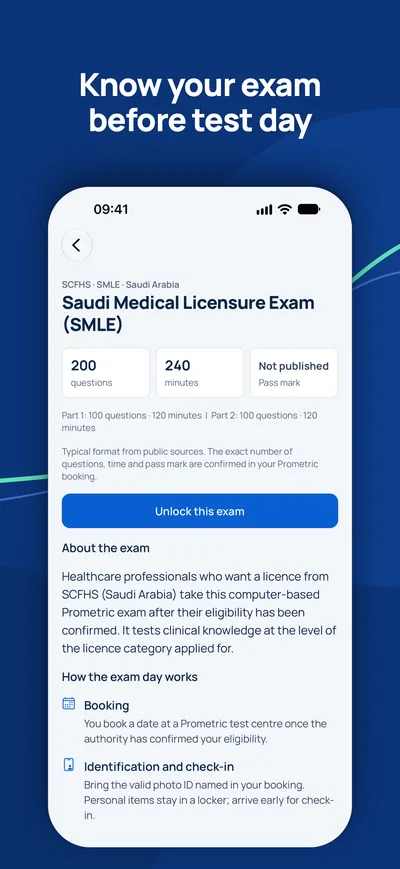
nicht veröffentlicht. Genaue Fragenzahl, Zeit und Bestehensgrenze stehen in Ihrer Buchung.
Welche Themen haben das größte Gewicht?+
Die größten Themen sind Zysten & Tumoren (20 %), Extraktion & Retention (15 %), Infektionen (15 %). Der Kurs umfasst 6 Themen mit insgesamt 30 Kapiteln.
Wie bereite ich mich auf die OMSB Fachzahnarzt Mund-, Kiefer- und Gesichtschirurgie-Prüfung vor?+
Arbeiten Sie die Kapitel Thema für Thema durch, wiederholen Sie die Lernnotizen und üben Sie dann mit zeitgesteuerten Probeprüfungen im Format Ihrer Behörde.
Fragen
Wie lange habe ich Zugang?+
Der Zugang gilt 90 Tage ab Zahlung für das gewählte Land und den gewählten Beruf.
Welche Geräte kann ich nutzen?+
Jeden Browser auf Computer, Tablet oder Handy sowie die MediFlow-App mit demselben Konto.
Kann ich das Geld zurückbekommen?+
Bitte lesen Sie vor dem Kauf unsere Erstattungsrichtlinie. Erstattungsrichtlinie
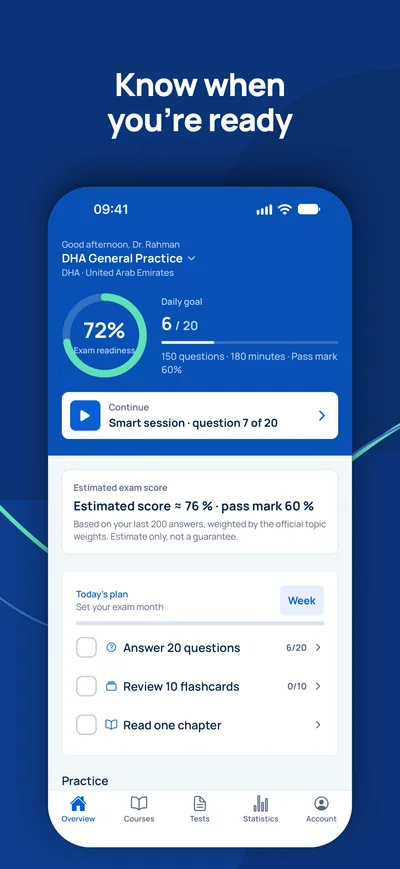
iPhone · iPad
GCC Medical Exam Prep
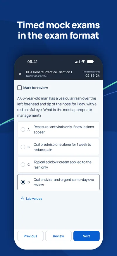
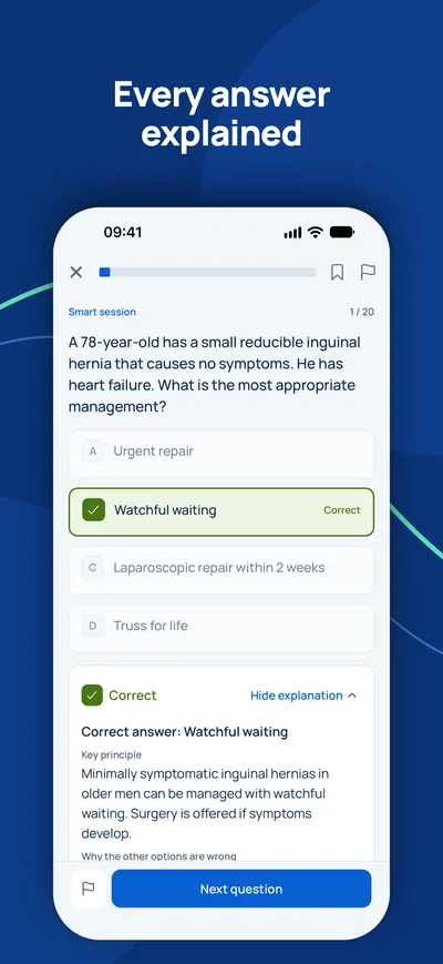
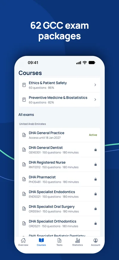
Bereiten Sie sich auf Ihrem iPhone auf DHA, DOH, MOHAP, SCFHS und weitere Zulassungsprüfungen am Golf vor.
Ist die Prüfung Ihr einziger offener Schritt?
Prüfen Sie zuerst, ob Sie die Prüfung überhaupt brauchen und welchen Titel Sie erwarten können. Ein Berater beantwortet Ihre Fragen kostenlos.
Eignungs-Check- 1Kostenlose Beratung per Video, Telefon oder WhatsApp
- 2Eignungs-Check gegen die offiziellen Anforderungen
- 3Ihr Plan mit Festpreisen und klaren nächsten Schritten