
🇴🇲 OMSB
Fachzahnarzt Prothetik Oman
Fachzahnärztinnen und Fachzahnärzte in der Vorbereitung auf die Prüfungen in Prothetik bei DHA, DOH, Qatar DHP und SCFHS

Beispielfrage
Which total occlusal convergence is regarded as a realistic clinical target for a full-crown preparation?
Keys: 1–5 or A–E to choose, Enter to check and continue, F to mark.
Original MediFlow content, not official exam questions. Medical review in progress.
Syllabus and high-yield notes
1. Festsitzend · 20 % Anteil in der Prüfung · 5 lessons+
- Eine Gesamtkonvergenz von 10–20° ist das klinisch realistische Ziel für Kronenpräparationen; die Retention sinkt mit zunehmender Konvergenz deutlich, kurze Wände brauchen Rillen, Kästen oder adhäsive Befestigung.
- Ränder, die in das suprakrestale Attachment (früher 'biologische Breite') eingreifen, verursachen chronische Entzündung oder Rezession. Etwa 3 mm vom geplanten Rand zum Knochenkamm einhalten, sonst chirurgische Kronenverlängerung oder Extrusion.
- Zirkonoxid-Brücken brauchen größere Verbinder als Metall: posterior gelten für 3Y-TZP etwa 9 mm² als Minimum. Lithiumdisilikat-Brücken sind auf drei Glieder bis höchstens zum zweiten Prämolaren begrenzt, mit Verbindern von etwa 16 mm².
- Einflügelige Adhäsivbrücken lösen sich seltener als zweiflügelige, weil unterschiedliche Pfeilerbewegungen die Klebung nicht mehr belasten.
- Die sofortige Dentinversiegelung (Adhäsiv auf frisch präpariertes Dentin vor der Abformung) verbessert die Haftung und verringert Hypersensibilitäten unter adhäsiven indirekten Restaurationen.
- Bei einem Zwischenpfeiler liegt die Patrize am Mesialen des distalen Brückenglieds und die Matrize am Distalen der Zwischenpfeilerkrone; bei Mesialbewegung setzt sich das Geschiebe fester.
- Sekundärkaries am Pfeiler ist die häufigste biologische Komplikation zahngetragener Brücken, gefolgt von endodontischem Behandlungsbedarf und Retentionsverlust.
- Lokaler Frontzahnverschleiß ohne interokklusalen Platz lässt sich nach dem Dahl-Konzept versorgen: Restaurationen in lokal erhöhter Vertikaldimension, danach stellen sich die Seitenzähne über Monate wieder in Kontakt.
- Abutments, biology and tooth preparation – Fixed prosthodontics replaces or restores teeth with restorations the patient cannot remove: crowns, inlays and onlays, veneers and fixed dental prostheses (FDPs, the current term for bridges). Every fixed restoration rests on three foundations. The abutment must be biologically able to carry load, the margin must respect the periodontium, and the preparation must give the restoration enough retention, resistance and material thickness. This chapter covers all three, because most clinical failures and most exam questions trace back to one of them.
- Materials, design and the fixed dental prosthesis – Choosing a material for a crown or FDP means matching strength, available space, aesthetics, the patient's function and the luting method. Modern practice offers three broad families: metal and metal-ceramic, glass ceramics (feldspathic, leucite-reinforced and lithium disilicate) and polycrystalline ceramics (zirconia). Each family has typical minimum thicknesses and connector sizes, and examiners test these numbers.
- Aesthetics, cementation, occlusion and complications – The final chapter follows the restoration from shade selection to the long-term recall. It covers how to choose shade, how to plan veneers so that bonding is reliable, how to lute different materials, how to transfer occlusion, how to treat localised wear conservatively, and what goes wrong over time.
- Aesthetic analysis and treatment of the anterior teeth – Aesthetic dentistry is a planned discipline, not a matter of taste. The prosthodontist analyses the face, the lips, the gingiva and the teeth in a fixed order, sets goals with the patient, tests them in the mouth with a mock-up and only then prepares teeth. The order runs from outside to inside: face, then smile, then gingiva, then teeth. This chapter gives the reference values that guide that analysis and the least invasive ways to reach the goal.
- Patient safety, ethics and professionalism in prosthodontics – Prosthodontic treatment is elective, often expensive, usually irreversible and frequently involves many teeth over many visits. That combination creates specific safety and ethical risks: preparing the wrong tooth, dropping a crown into the airway, overtreating a patient for profit, or failing to explain the long-term costs of maintenance. The Qatar specialty blueprint gives patient safety and professionalism together 10 % of the prosthodontics examination. This chapter brings the principles together.
2. Teilprothetik · 15 % Anteil in der Prüfung · 5 lessons+
- Führungsflächen sind parallele approximale Flächen von etwa 2–4 mm Höhe; sie legen eine Einschubrichtung fest und tragen zu Stabilität und Reziprozität bei.
- Beim RPI-System liegt der I-Steg mesiobukkal unterhalb des prothetischen Äquators; bei Belastung der Freiendsattels bewegt er sich nach gingival und mesial und löst sich vom Zahn, was den Pfeiler schont.
- Der reziproke Arm muss den Zahn berühren, bevor die Retentionsspitze den Äquator passiert; er liegt daher auf oder oberhalb der Prothetikäquatorlinie der Gegenseite.
- Cingulumauflagen am Eckzahn sind Inzisalauflagen vorzuziehen: Die Kraft wirkt axialer, näher am Drehzentrum, und sie sind weniger sichtbar.
- Ein Implantat am distalen Ende einer Freiendsituation (Kennedy I) macht den Sattel zu einer zahn- und implantatgestützten Basis, verringert Rotation und verbessert die Abstützung (implantatunterstützte Teilprothese).
- Einseitige herausnehmbare Teilprothesen ohne Querstabilisierung sind wegen geringer Stabilität und Aspirationsgefahr kontraindiziert.
- Hebt sich bei Druck auf den Freiendsattel die indirekte Halterung von ihrer Auflage ab, hat der Sattel die Gewebeabstützung verloren und muss unterfüttert werden.
- Flexible Nylonprothesen haben keine starren Auflagen und sind schwer zu unterfüttern oder zu reparieren; sie eignen sich für den Übergang oder bei Methacrylat-Allergie, nicht als definitive Versorgung.
- Classification, biomechanics and the surveyor – A removable partial denture (RPD) replaces missing teeth in a partially dentate arch and can be taken out by the patient. Its design is a balance between support (resistance to movement towards the tissues), retention (resistance to movement away from the tissues) and stability (resistance to horizontal and rotational movement). To design well you must first describe the arch, then understand how the denture moves under load, then plan with a surveyor. This chapter covers those three steps.
- Components of the RPD: connectors, rests, clasps and guiding planes – Every metal-framework RPD is built from the same parts: a major connector, minor connectors, rests, direct retainers (clasps or attachments), indirect retainers, guiding planes and denture bases. Knowing what each part does and the rules for its design answers most RPD questions.
- Mouth preparation, special designs, delivery and maintenance – Once the design is drawn on the surveyed cast, the mouth is prepared to match it. Preparation proceeds in a set order, because each step depends on the previous one. Then the framework is tried in, the bases are added, the denture is delivered and the patient enters a lifelong recall. This chapter also covers designs for difficult situations: overdentures, swing-lock dentures, flexible dentures and the occlusion against a complete denture.
- Maxillofacial prosthodontics: maxillary defects, obturators and speech prostheses – Maxillofacial prosthodontics restores parts of the mouth and face lost through surgery, trauma or congenital defects. Most cases follow cancer surgery. The prosthodontist works within a team that includes the head and neck surgeon, radiation oncologist, speech and language therapist and dietitian. This chapter covers defects of the maxilla and soft palate. The next chapter covers the cancer patient more broadly, mandibular defects and facial prostheses.
- The head and neck cancer patient, mandibular defects and facial prostheses – Patients treated for head and neck cancer face a lifetime of oral problems. Radiotherapy damages salivary glands, blood vessels, muscles and bone. Surgery removes parts of the jaws, tongue and face. The prosthodontist helps prepare the mouth before treatment, prevents complications during and after it and restores function and appearance. This chapter covers those stages, mandibular resection and facial prostheses.
3. Totalprothetik · 15 % Anteil in der Prüfung · 5 lessons+
- Der ACP Prosthodontic Diagnostic Index stuft die Unterkieferknochenhöhe ein: Klasse I mindestens 21 mm, Klasse II 16–20 mm, Klasse III 11–15 mm, Klasse IV 10 mm oder weniger.
- Die neutrale Zone ist der Raum, in dem sich Zungen- und Wangen-/Lippenkräfte ausgleichen; ihre Registrierung ist Methode der Wahl bei instabiler Unterkieferprothese auf stark atrophiertem Kiefer.
- Die Okklusionsebene im Unterkiefer verläuft vom Eckzahn nach dorsal bis etwa zur Hälfte bis zu zwei Dritteln der Höhe des retromolaren Polsters.
- Bei der lingualisierten Okklusion stehen die palatinalen Höcker der Oberkieferzähne in den zentralen Gruben der Unterkieferzähne ohne bukkale Kontakte; sie eignet sich bei atrophiertem Unterkiefer und hält die Kräfte lingual.
- Generalisierte Druckstellen und eine nach vorn rutschende Unterkieferprothese weisen meist auf einen okklusalen Fehler in zentrischer Relation hin; eine klinische Remontage korrigiert dies am genauesten.
- Miconazol ist unter Warfarin kontraindiziert, weil es den INR erhöht; Nystatin ist bei antikoagulierten Patienten das sicherere Antimykotikum bei Prothesenstomatitis.
- Kopierprothesen übernehmen polierte Flächen und Zahnstellung gut angenommener alter Prothesen und erleichtern älteren Menschen die Eingewöhnung.
- Das Christensen-Phänomen ist das dorsale Auseinanderklaffen der Bisswälle oder Zähne bei Protrusion durch die nach kaudal gerichtete Kondylenbahn; Kompensationskurven ermöglichen balancierte Kontakte.
- The edentulous patient: anatomy, classification and impressions – Complete dentures replace all teeth in an arch and rest entirely on mucosa over bone. They have no teeth to hold onto, so success depends on how well the dentist understands the tissues that support them, the muscles that move around them and the patient who has to learn to control them. This chapter covers the anatomy of the denture-bearing areas, how the difficulty of a case is classified, and how impressions capture the tissues.
- Jaw relations, tooth arrangement and occlusion – With the impressions done, the dentist must decide where the teeth will go. This requires three decisions: the vertical dimension, the horizontal jaw relation (centric relation) and the position of the teeth and occlusal plane. Then an occlusal scheme is chosen. Errors here cause most problems patients later blame on the 'fit' of the denture.
- Insertion, problems, special techniques and denture-related disease – Delivering complete dentures is the start of a period of adaptation, not the end of treatment. The dentist checks fit, extension and occlusion, teaches the patient, and then works through the problems that arise in the first weeks. This chapter covers insertion, processing faults, common complaints and their causes, special techniques for difficult patients, and the diseases associated with denture wearing.
- Overdentures, immediate dentures, copy dentures and digital workflows – The conventional complete denture is made on healed ridges after all teeth are gone. Many patients do not fit that pattern. Some still have a few teeth or roots that can be kept, some need teeth removed and want to leave the surgery with a denture, some are old and frail and have adapted to a worn denture they will not give up, and some can be treated with digitally designed and milled dentures. This chapter explains the logic of each option, when it is indicated and what goes wrong.
- The older and medically compromised edentulous patient – Most complete denture wearers are older and many take several medicines. Their general health changes the oral tissues, the ability to adapt and the risks of treatment. Prosthodontic success therefore depends on medical history as much as on technique. This chapter covers the conditions examiners ask about most: xerostomia, candidiasis and denture stomatitis, neurological disease, dementia, anticoagulation, antiresorptive drugs, radiotherapy, nutrition and safe use of denture adhesives.
4. Implantatprothetik · 20 % Anteil in der Prüfung · 5 lessons+
- Falldefinition Periimplantitis 2017: Blutung und/oder Suppuration bei sanfter Sondierung, zunehmende Sondierungstiefe und Knochenverlust über das initiale Remodelling hinaus; ohne Ausgangsbefund Knochenniveau mindestens 3 mm und/oder Sondierungstiefe mindestens 6 mm mit Blutung.
- Die EFP-S3-Leitlinie (2023) empfiehlt bei Periimplantitis zuerst nichtchirurgische Therapie, bei Bedarf danach chirurgische; systemische Antibiotika als Zusatz werden nicht routinemäßig empfohlen.
- In der ästhetischen Zone liegt die Implantatschulter etwa 3–4 mm apikal des geplanten bukkalen Gingivarands, leicht palatinal, mit mindestens 2 mm bukkalem Knochen.
- Zwischen zwei benachbarten Implantaten ist die Papille im Mittel nur etwa 3–4 mm hoch; benachbarte Implantate in der ästhetischen Zone sollten möglichst vermieden werden.
- Zementüberschuss ist eine häufige Ursache periimplantärer Erkrankungen; bei tiefen Rändern ist Verschraubung oder ein Rand auf Höhe der Mukosa vorzuziehen.
- Bei Patienten unter hochdosierter intravenöser antiresorptiver Therapie wegen einer Krebserkrankung sind Implantate wegen des hohen MRONJ-Risikos kontraindiziert.
- Implantatgeschützte Okklusion: leichter oder kein Kontakt bei leichtem Biss, gleichmäßiger Kontakt bei festem Biss, möglichst keine Kontakte bei Exkursionen, schmale Kauflächen und zentrale Belastung.
- Kurze Implantate (6 mm oder weniger) im atrophierten Seitenzahnbereich des Unterkiefers erreichen vergleichbare Überlebensraten wie längere Implantate nach vertikaler Augmentation, bei weniger Komplikationen.
- Patient selection, risk and treatment planning for implants – An implant is a titanium or zirconia root analogue that becomes anchored in bone by osseointegration. Osseointegration means direct structural and functional contact between living bone and the implant surface, without an intervening soft-tissue layer. Unlike a natural tooth, an implant has no periodontal ligament. This single fact explains much of implant prosthodontics: an implant cannot move physiologically, gives little proprioceptive feedback, cannot migrate to relieve load, and has a connective tissue seal that runs parallel to the surface instead of inserting into it. The prosthodontist plans the restoration first and places the implant where the restoration needs it. This is called restoration-driven or prosthetically driven planning.
- The aesthetic zone, soft tissue and implant components – In the anterior maxilla success is judged not only by survival but by the appearance of the mucosa. The goal is a mid-facial margin at the level of the contralateral tooth, papillae filling the embrasures and no grey shine-through. These depend on three-dimensional implant position, bone and soft-tissue volume, and the restorative components.
- Biomechanics, occlusion and multi-unit implant prostheses – Because implants lack a periodontal ligament, forces are transmitted directly to bone, mostly at the crest. A natural tooth intrudes about 25–100 micrometres under load; an implant moves only about 3–5 micrometres. This mismatch, together with reduced tactile sensitivity, guides how we design occlusion and frameworks. Overload rarely causes bone loss in a healthy, plaque-free site, but it is a major cause of technical complications: screw loosening, abutment fracture, veneer chipping and, rarely, implant fracture.
- Peri-implant diseases: diagnosis, treatment and maintenance – Peri-implant health, mucositis and peri-implantitis were defined at the 2017 World Workshop. Peri-implant mucositis is inflammation of the soft tissue only: bleeding and/or suppuration on gentle probing, with or without increased probing depth, but no bone loss beyond the initial remodelling that occurs in the first year. It is reversible with plaque control. Peri-implantitis is a plaque-associated pathological condition with inflammation of the mucosa and progressive loss of supporting bone.
- Surgical complications, mishaps and the evidence for implant decisions – Implant treatment has high survival rates, but complications are common enough that every clinician must prevent, recognise and manage them. Complications are usually grouped by time: during surgery, early (before osseointegration) and late (after loading). They are also grouped by type: biological, technical and aesthetic. This chapter covers surgical mishaps, early failure, technical problems and the evidence that should guide treatment choices.
5. Okklusion & CMD · 15 % Anteil in der Prüfung · 5 lessons+
- DC/TMD Achse I: Myalgie erfordert Schmerzen der Kaumuskulatur, die durch Kieferfunktion beeinflusst werden, und bekannten Schmerz bei Palpation oder Öffnung; myofaszialer Schmerz mit Übertragung zeigt Schmerz über den palpierten Muskel hinaus.
- Diskusverlagerung ohne Reposition mit eingeschränkter Öffnung: Anamnese einer Kiefersperre, maximale passive Öffnung unter 40 mm einschließlich Überbiss, Deflexion zur betroffenen Seite.
- Der Bruxismus-Konsens 2018 unterscheidet möglich (Selbstangabe), wahrscheinlich (Selbstangabe plus klinischer Befund) und sicher (instrumentelle Messung, Polysomnographie beim Schlafbruxismus).
- Eine Stabilisierungsschiene ist eine harte Vollbogenschiene mit gleichmäßigen Kontakten in zentrischer Relation und Eckzahnführung; irreversible Einschleiftherapie wird zur Behandlung von CMD nicht empfohlen.
- Steilere Kondylen- und Frontzahnführung erlauben höhere Seitenzahnhöcker; ein größerer Immediate Side Shift erfordert flachere Höcker und weitere Fossae.
- Gegenseitig geschützte Okklusion: Die Seitenzähne schützen die Frontzähne in maximaler Interkuspidation, die Frontzähne disokkludieren die Seitenzähne bei Exkursionen.
- Die Magnetresonanztomographie zeigt Diskusposition und Gelenkerguss; die DVT zeigt knöcherne Veränderungen wie Abflachung, Erosion, Osteophyten und subchondrale Zysten.
- Chronische CMD mit ausgedehnten Schmerzen, Schlafstörungen und Belastung weisen auf zentrale Sensibilisierung hin und erfordern ein biopsychosoziales Vorgehen: Aufklärung, Selbstmanagement, kognitive Verhaltenstherapie und bei Bedarf niedrig dosierte trizyklische Antidepressiva.
- Functional anatomy of the TMJ and mandibular movement – The temporomandibular joint (TMJ) is a ginglymoarthrodial joint: it hinges (rotation) and glides (translation). The mandibular condyle sits in the glenoid (mandibular) fossa of the temporal bone, in front of which lies the articular eminence. Between them is the articular disc, a biconcave plate of dense fibrous connective tissue without vessels or nerves in its central intermediate zone. The disc has a thick posterior band, a thin intermediate zone and a thinner anterior band. In the normal closed position the posterior band sits at about the 12 o'clock position on top of the condyle.
- Occlusal concepts and determinants for restorative work – An occlusal scheme is the planned pattern of tooth contact in closure and in excursions. For dentate patients the reference concept is mutually protected occlusion: in maximum intercuspation, the posterior teeth carry the vertical load and protect the anterior teeth; in protrusive and lateral excursions, the anterior teeth (incisors and canines) guide the mandible and immediately disclude the posterior teeth, protecting them from horizontal forces. Posterior teeth tolerate axial loads well; anterior teeth have a favourable position far from the muscles and good proprioception to manage lateral guidance.
- Temporomandibular disorders and bruxism: diagnosis – Temporomandibular disorders (TMD) are a group of musculoskeletal and neuromuscular conditions affecting the masticatory muscles, the TMJs and associated structures. They are the most common cause of non-dental orofacial pain. Diagnosis follows the Diagnostic Criteria for TMD (DC/TMD, Schiffman 2014), which has two axes. Axis I is the physical diagnosis based on history and a standardised examination. Axis II screens psychosocial status and pain-related disability (for example the Graded Chronic Pain Scale, PHQ-4 for depression and anxiety, jaw function limitation scales). Both axes matter, because psychosocial factors predict chronicity.
- Management of TMD: splints, conservative care and chronic pain – Most TMD is self-limiting or fluctuating, and conservative, reversible treatment helps most patients. Irreversible treatments such as occlusal adjustment by selective grinding, orthodontics or prosthodontic reconstruction aimed at curing TMD are not supported by evidence and are not recommended. The occlusion is not the main cause of TMD, and changing it permanently may create new problems.
- The worn dentition and full-mouth rehabilitation – Tooth wear is the loss of hard tissue by processes other than caries or trauma. It is common, increases with age and is becoming more frequent in young people because of acidic drinks and reflux. Most wear is slow and needs only monitoring. Severe or rapid wear, however, can lead to sensitivity, poor appearance, fractures and loss of crown height, and may need complex rehabilitation. This chapter covers diagnosis, monitoring and the principles of restoring a worn dentition, including how to manage vertical dimension and choose the reference position.
6. Werkstoffe · 15 % Anteil in der Prüfung · 5 lessons+
- 5Y-Zirkonoxid (mit kubischem Anteil) ist transluzenter als 3Y-TZP, hat aber geringere Biegefestigkeit und kaum Umwandlungsverstärkung; es braucht dickere Wandstärken und größere Verbinder.
- Die Niedertemperaturdegradation von Zirkonoxid ist eine langsame tetragonal-monokline Umwandlung in feuchter Umgebung, die die Oberfläche aufraut und die Festigkeit mindern kann.
- Gracis 2015 teilt Keramiken in glasmatrixbasierte (Feldspat, Leuzit, Lithiumdisilikat), polykristalline (Aluminiumoxid, Zirkonoxid) und kunststoffmatrixbasierte (Nanokeramik, polymerinfiltriertes Keramiknetzwerk) ein.
- Polierte monolithische Zirkonoxid-Oberflächen verursachen weniger Schmelzabrasion am Antagonisten als glasierte Zirkonoxid- oder Verblendkeramik; eingeschliffenes Zirkonoxid muss sorgfältig poliert werden.
- Polyether ist steif und hydrophil und nimmt Wasser auf; Abformungen müssen trocken gelagert werden. Für Implantat-Pick-up-Abformungen gut geeignet.
- Intraoralscanner sind bei kurzen Spannen so genau wie konventionelle Abformungen; über den ganzen Kiefer nehmen die Fehler zu, besonders bei mehreren Implantaten.
- Titangüsse bilden eine Alpha-Case-Schicht, eine harte, spröde, sauerstoffangereicherte Oberflächenschicht durch Reaktion mit der Einbettmasse, die entfernt werden muss.
- Gedruckte Kunststoffe für Prothesen und Schienen müssen nach Herstellervorgabe gereinigt und nachgehärtet werden, sonst bleiben Restmonomer und schlechte mechanische Eigenschaften.
- Dental ceramics: classification, zirconia and material choice – Ceramics are inorganic, non-metallic materials. In the mouth they offer aesthetics, biocompatibility and chemical stability, but they are brittle: they have high compressive strength, lower tensile strength and limited ability to resist crack growth. Two properties guide clinical choice. Flexural strength (MPa) describes the stress at which a test bar fractures. Fracture toughness (MPa·m^1/2, K_IC) describes resistance to growth of an existing crack, and for brittle materials it is often the more meaningful number, because failures start from flaws.
- Impressions, digital workflow, gypsum and resins – Precision of the final restoration begins with the impression. Elastomeric impression materials used in prosthodontics are addition silicone (polyvinyl siloxane, PVS), polyether, condensation silicone and polysulphide. Their key properties are accuracy, dimensional stability, elastic recovery, tear strength, stiffness and wettability (hydrophilicity).
- Metals, alloys, titanium and resin bonding – Dental casting alloys are classified (ADA) by noble metal content: high noble (at least 60 % noble metal with at least 40 % gold), noble (at least 25 % noble metal), and predominantly base metal (less than 25 % noble). Noble metals are gold, platinum, palladium and others of the platinum group; silver is not counted as noble in this classification. Base metal alloys such as cobalt-chromium and nickel-chromium are stiffer (high elastic modulus), harder and cheaper, which suits thin long-span frameworks and removable partial denture frameworks, but they are harder to cast and adjust. Nickel can cause allergy, especially in women.
- Luting cements and adhesive cementation – A luting cement fills the gap between a restoration and the prepared tooth or abutment. It must flow into a thin film, set in a controlled time, resist dissolution, and, depending on type, either rely on mechanical interlocking or bond chemically or micromechanically. The choice depends on the retention and resistance form of the preparation, the restoration material, the ability to isolate the field and the patient's caries risk. This chapter compares the main cement families and explains the bonding protocols for different substrates.
- Physical and mechanical properties, biocompatibility and material testing – To understand why a ceramic chips, why a denture base cracks or why a clasp fractures, you need the basic language of material science. This chapter defines the properties examiners ask about, explains how they are tested and links each property to a clinical decision. It then covers biocompatibility, allergy and the standards that govern dental materials.
Festsitzend
Lesson 1Abutments, biology and tooth preparation
Fixed prosthodontics replaces or restores teeth with restorations the patient cannot remove: crowns, inlays and onlays, veneers and fixed dental prostheses (FDPs, the current term for bridges). Every fixed restoration rests on three foundations. The abutment must be biologically able to carry load, the margin must respect the periodontium, and the preparation must give the restoration enough retention, resistance and material thickness. This chapter covers all three, because most clinical failures and most exam questions trace back to one of them.
An abutment is a tooth (or implant) that supports and retains an FDP. Its value depends on the crown-root ratio, the root configuration and the periodontal surface area. The crown-root ratio compares the length of tooth above the alveolar crest with the length of root in bone. An ideal ratio is 1:2; 2:3 is the realistic optimum; 1:1 is the minimum acceptable for an abutment under normal occlusal load. As bone is lost, the clinical crown lengthens, the lever arm grows and the tooth becomes less able to resist lateral forces.
Root form matters as much as length. Roots that are wider labiolingually than mesiodistally resist rotation better than round roots. Multirooted teeth with widely separated roots spread forces over a larger area of bone and are the most favourable abutments. Fused, conical or tapered roots behave like a single root and give less support. Irregular curvature or dilaceration helps a little more than a perfectly conical root. Ante's law, an older guideline, states that the combined root surface area of the abutments should be at least equal to that of the teeth being replaced. It is a useful reminder of load sharing but is not supported as a strict rule; long spans fail mainly through flexure of the FDP and through biology rather than through root area alone.
| Factor | Favourable | Unfavourable |
|---|---|---|
| Crown-root ratio | 2:3 or better | Worse than 1:1 |
| Root configuration | Widely separated roots; broad labiolingually | Fused or conical roots |
| Periodontal status | Stable, no mobility progression | Uncontrolled periodontitis, increasing mobility |
| Span length | Single pontic | Long spans (flexure rises with the cube of the span length) |
| Arch form | Straight span | Curved anterior span crossing the inter-abutment axis |
Teeth with a compromised root can sometimes still be used. A mandibular molar with a through-and-through (degree III) furcation can be hemisected: the crown and root are split, the hopeless root is extracted and the other half is treated endodontically and used as a premolar-sized abutment. In the mandibular molar the distal root is usually larger, straighter and easier to treat than the mesial root, which has two canals and a curvature. In the maxilla the same idea is called root resection or trisection. Such teeth need endodontic treatment, a short span, cleansable embrasures and a patient who accepts a guarded prognosis.
Margins must respect the supracrestal tissue attachment. This term (2017 World Workshop classification) replaces 'biologic width'. It is the junctional epithelium plus the connective tissue attachment coronal to the alveolar crest, together roughly 2 mm. Add a healthy sulcus of about 1 mm and you reach the clinical rule: keep about 3 mm between the planned restoration margin and the bone crest. A margin placed inside this zone produces chronic inflammation, bleeding and pocketing in a thick biotype, or recession and bone loss in a thin biotype. The tissue tries to re-establish its dimension at the expense of bone.
The violation is diagnosed by bone sounding: under local anaesthesia a periodontal probe is pushed through the soft tissue to the bone. If the distance from margin to crest is under about 3 mm and the gingiva stays inflamed despite good hygiene and a good fit, the margin is too deep. Replacing the crown does not help. Management is surgical crown lengthening with ostectomy to re-create about 3 mm, or orthodontic extrusion (with fiberotomy if the bone should not follow) when aesthetics forbid removing bone around one anterior tooth. Extrusion also keeps the gingival level of neighbouring teeth intact.
When a proximal box ends only slightly below the cemento-enamel junction and the attachment is intact, surgery is not needed. Deep margin elevation places a layer of composite in the deep part of the box under rubber dam or a matrix band, moving the margin coronally. The indirect restoration is then made on this elevated base, which simplifies impression or scanning and adhesive cementation. It is only acceptable when the supracrestal attachment is not violated and isolation is reliable.
Preparation design follows five principles: preservation of tooth structure, retention and resistance form, structural durability (enough thickness for the material), marginal integrity and preservation of the periodontium. Retention prevents removal along the path of insertion; resistance prevents dislodgement by oblique forces. Both rise when the axial walls are nearly parallel, long and close together. Total occlusal convergence (TOC) is the angle between two opposing axial walls. Laboratory retention is greatest at about 6°, but studies of real preparations show that clinicians achieve much more. A target of 10 to 20° is accepted as realistic. Above about 20° retention falls sharply.
| Restoration | Occlusal reduction | Axial reduction | Margin |
|---|---|---|---|
| Full metal (gold) crown | About 1.5 mm functional, 1.0 mm non-functional cusps | About 0.5 mm | Light chamfer |
| Metal-ceramic crown | About 2.0 mm functional, 1.5 mm non-functional cusps | 1.2–1.5 mm facially | Shoulder or heavy chamfer facially, chamfer lingually |
| Monolithic lithium disilicate crown | About 1.5 mm | About 1.0–1.5 mm | Rounded shoulder or deep chamfer about 1.0 mm |
| Monolithic zirconia (3Y-TZP) crown | About 0.5–1.0 mm (manufacturer dependent) | About 0.5–1.0 mm | Chamfer |
Sources: Rosenstiel SF, Land MF, Walter R. Contemporary Fixed Prosthodontics, 6th ed. Elsevier, 2023. · Shillingburg HT, Sather DA, Wilson EL, et al. Fundamentals of Fixed Prosthodontics, 4th ed. Quintessence, 2012. · Goodacre CJ, Campagni WV, Aquilino SA. Tooth preparations for complete crowns: an art form based on scientific principles. J Prosthet Dent 2001;85:363–376. · Jepsen S, Caton JG, et al. Periodontal manifestations of systemic diseases and developmental and acquired conditions: consensus report of workgroup 3 of the 2017 World Workshop. J Clin Periodontol 2018;45(Suppl 20):S219–S229.
Zur OMSB Fachzahnarzt Prothetik-Prüfung
Wie viele Fragen hat die OMSB Fachzahnarzt Prothetik-Prüfung?+
nicht veröffentlicht. Genaue Fragenzahl, Zeit und Bestehensgrenze stehen in Ihrer Buchung.
Welche Themen haben das größte Gewicht?+
Die größten Themen sind Festsitzend (20 %), Implantatprothetik (20 %), Teilprothetik (15 %). Der Kurs umfasst 6 Themen mit insgesamt 30 Kapiteln.
Wie bereite ich mich auf die OMSB Fachzahnarzt Prothetik-Prüfung vor?+
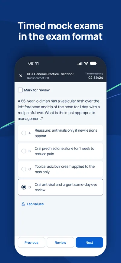
Arbeiten Sie die Kapitel Thema für Thema durch, wiederholen Sie die Lernnotizen und üben Sie dann mit zeitgesteuerten Probeprüfungen im Format Ihrer Behörde.
Fragen
Wie lange habe ich Zugang?+
Der Zugang gilt 90 Tage ab Zahlung für das gewählte Land und den gewählten Beruf.
Welche Geräte kann ich nutzen?+
Jeden Browser auf Computer, Tablet oder Handy sowie die MediFlow-App mit demselben Konto.
Kann ich das Geld zurückbekommen?+
Bitte lesen Sie vor dem Kauf unsere Erstattungsrichtlinie. Erstattungsrichtlinie

iPhone · iPad
GCC Medical Exam Prep
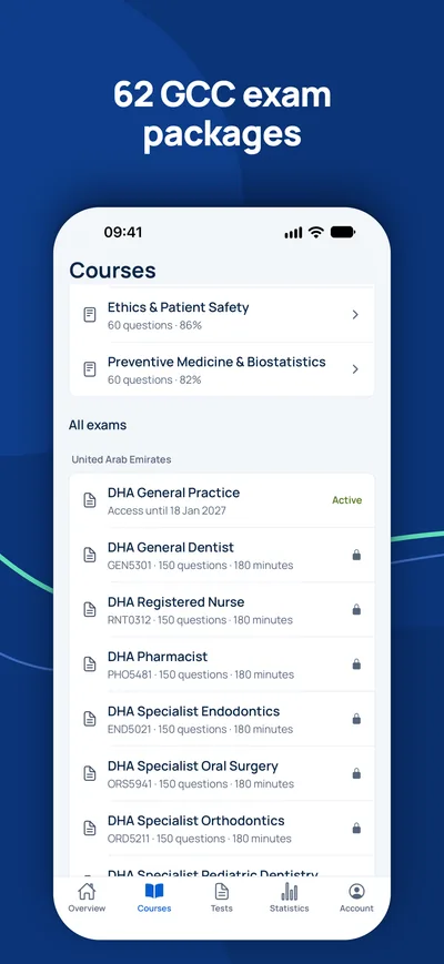
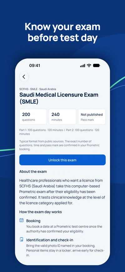
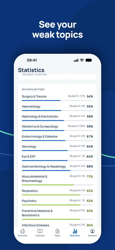
Bereiten Sie sich auf Ihrem iPhone auf DHA, DOH, MOHAP, SCFHS und weitere Zulassungsprüfungen am Golf vor.


Ist die Prüfung Ihr einziger offener Schritt?
Prüfen Sie zuerst, ob Sie die Prüfung überhaupt brauchen und welchen Titel Sie erwarten können. Ein Berater beantwortet Ihre Fragen kostenlos.
Eignungs-Check- 1Kostenlose Beratung per Video, Telefon oder WhatsApp
- 2Eignungs-Check gegen die offiziellen Anforderungen
- 3Ihr Plan mit Festpreisen und klaren nächsten Schritten