
🇶🇦 DHP
Fachzahnarzt Parodontologie Katar
Fachzahnärztinnen und Fachzahnärzte in der Vorbereitung auf die Prüfungen in Parodontologie bei DHA, DOH, Qatar DHP und SCFHS

Beispielfrage
Which drug class is NOT typically associated with drug-influenced gingival enlargement?
Keys: 1–5 or A–E to choose, Enter to check and continue, F to mark.
Original MediFlow content, not official exam questions. Medical review in progress.
Syllabus and high-yield notes
1. Klassifikation & Diagnostik · 17 % Anteil in der Prüfung · 5 lessons+
- Das Staging (I–IV) beschreibt Schweregrad und Komplexität: interdentaler CAL an der schlechtesten Stelle, röntgenologischer Knochenabbau, parodontal bedingter Zahnverlust und Komplexitätsfaktoren (ST ≥6 mm, vertikale Defekte ≥3 mm, Furkation II/III, Kaufunktionsstörung, weniger als 20 Zähne). Komplexitätsfaktoren können das Stadium nur anheben.
- Der Grad (A–C) richtet sich primär nach direktem Progressionsnachweis, sonst nach %Knochenabbau/Alter am schlechtesten Zahn (<0,25 A, 0,25–1,0 B, >1,0 C); Grad B ist Standard und wird durch Rauchen (≥10 Zig./Tag = C) und HbA1c (≥7,0 % = C) modifiziert.
- Ausdehnung: lokalisiert bei weniger als 30 % der Zähne, generalisiert ab 30 %; das Molaren-Inzisiven-Muster wird gesondert beschrieben und ersetzt die frühere lokalisierte aggressive Parodontitis.
- Ein „Parodontitisfall“ liegt vor bei interdentalem CAL an ≥2 nicht benachbarten Zähnen oder bukkalem/oralem CAL ≥3 mm mit ST >3 mm an ≥2 Zähnen, sofern nicht anders erklärbar.
- Gingivitis am intakten Parodont: BOP ≥10 % bei ST ≤3 mm; lokalisiert 10–30 %, generalisiert >30 %. Ein erfolgreich behandelter Parodontitispatient mit BOP <10 % ohne ST ≥4 mm mit BOP ist „parodontal gesund am reduzierten Parodont“, bleibt aber Parodontitispatient.
- Nekrotisierende Parodontalerkrankungen werden nach Wirtsstatus eingeteilt: chronisch schwer kompromittiert (z. B. HIV/AIDS, schwere Mangelernährung) oder vorübergehend bzw. mäßig kompromittiert (z. B. Stress, Rauchen, Schlafmangel).
- Gingivarezessionen werden nach interdentalem CAL eingeteilt (Cairo RT1–RT3), was die erreichbare Wurzeldeckung vorhersagt: RT1 kein approximaler Verlust, RT2 approximaler CAL ≤ bukkal, RT3 approximaler CAL > bukkal.
- Endo-parodontale Läsionen werden seit 2017 nach Vorhandensein einer Wurzelschädigung (Fraktur, Perforation, externe Resorption) und ohne Schädigung nach Parodontitisstatus und Ausdehnung der tiefen Tasche (schmal, breit, eine oder mehrere Flächen) eingeteilt.
- Periodontal health, gingivitis and the logic of the 2017 classification – In 2017 the American Academy of Periodontology (AAP) and the European Federation of Periodontology (EFP) held a World Workshop that replaced the 1999 Armitage classification. The 2018 consensus papers are now the language of every Gulf licensing exam. The biggest changes were three. First, periodontal health was defined for the first time in measurable terms. Second, the old split into chronic and aggressive periodontitis was dropped, because the two could not be separated reliably by biology. Third, every periodontitis case is now described by a stage (how severe and how complex) and a grade (how fast it progresses and how much risk the patient carries).
- Staging and grading of periodontitis step by step – Periodontitis is chronic inflammation, driven by a dysbiotic biofilm, that destroys the attachment apparatus. A patient is a periodontitis case when interdental CAL is detectable at two or more non-adjacent teeth, or when buccal or oral CAL of 3 mm or more with pocketing of more than 3 mm is present at two or more teeth, and the loss cannot be explained by other causes such as traumatic recession, caries at the cervical area, a vertical root fracture, an endodontic lesion draining through the periodontium or a third molar extraction.
- Necrotizing disease, systemic conditions, abscesses, endo-perio lesions and mucogingival conditions – Necrotizing periodontal diseases are a separate category of periodontitis because their course is acute and their link to impaired host defence is strong. Necrotizing gingivitis (NG) affects only the gingiva; necrotizing periodontitis (NP) also destroys attachment and bone; necrotizing stomatitis spreads beyond the gingiva into the mucosa and bone. The triad for diagnosis is necrosis and ulceration of interdental papillae (punched-out, often covered by a grey pseudomembrane), gingival bleeding and pain. Fetid breath, fever and lymphadenopathy are supportive. Predisposing factors include psychological stress, sleep deprivation, smoking, malnutrition and immunosuppression, especially HIV infection with low CD4 counts. The classification separates patients who are chronically severely compromised from those who are temporarily or moderately compromised, such as a student during exams.
- Periodontal structures: gingiva, periodontal ligament, cementum and alveolar bone – The periodontium is the group of tissues that attach the tooth to the jaw and protect that attachment. It has four parts: the gingiva, the periodontal ligament (PDL), the root cementum and the alveolar bone. The gingiva is the only part you see clinically; the other three are judged with the probe and the radiograph. Every diagnosis in periodontics is a statement about one or more of these tissues, so knowing their normal structure is the base for everything else in this track.
- Periodontal examination, charting and radiographic assessment – A periodontal diagnosis is only as good as the examination behind it. The classification in this topic needs specific numbers: interdental clinical attachment loss, radiographic bone loss in relation to root length, probing depths, furcation involvement, mobility, tooth loss due to periodontitis and bleeding on probing. This chapter explains how to collect those numbers correctly and how screening tools fit in.
2. Ätiologie & Risiko · 17 % Anteil in der Prüfung · 5 lessons+
- Aktuelles Pathogenesemodell: polymikrobielle Synergie und Dysbiose (PSD). Keystone-Pathogene wie Porphyromonas gingivalis unterwandern die Wirtsabwehr schon in geringer Menge, und die fehlregulierte Entzündung nährt die dysbiotische Gemeinschaft.
- Knochenabbau wird über das RANKL/OPG-Gleichgewicht gesteuert: Th17-Zellen, Osteoblasten und Fibroblasten exprimieren RANKL; IL-17, IL-1β, TNF-α und PGE2 verschieben das Verhältnis zugunsten von RANKL gegenüber Osteoprotegerin.
- Läsionen nach Page und Schroeder: initial (2–4 Tage, Neutrophile), früh (4–7 Tage, T-Lymphozyten), etabliert (2–3 Wochen, Plasmazellen/B-Zellen), fortgeschritten (Knochenabbau, Übergang zur Parodontitis).
- Rauchen erhöht das Parodontitisrisiko dosisabhängig (etwa 2- bis 7-fach), maskiert die Blutung durch Vasokonstriktion und verschlechtert die Heilung nach nichtchirurgischer und chirurgischer Therapie.
- Der Zusammenhang Diabetes–Parodontitis ist bidirektional: schlechte Einstellung erhöht das Parodontitisrisiko, und Parodontaltherapie senkt den HbA1c nach 3 Monaten um etwa 0,3–0,4 Prozentpunkte.
- Advanced Glycation End-Products (AGEs) binden an RAGE auf Makrophagen und Endothel, verstärken die Zytokinfreisetzung und stören den Kollagenumsatz bei Diabetikern.
- Lokale plaqueretentive Faktoren sind überstehende Restaurationen, subgingivale Ränder, Schmelzperlen, zervikale Schmelzausläufer und palatinale Rinnen; ihre Korrektur gehört zu Stufe 1 bzw. 2 der Therapie.
- Okklusales Trauma löst keine Parodontitis aus, kann aber bei plaquebedingter Entzündung durch Jiggling-Kräfte den Attachmentverlust beschleunigen (Glickman-Kodestruktionskonzept vs. Waerhaug).
- The biofilm and the microbiology of periodontitis – Dental plaque is a biofilm: a structured community of microorganisms attached to the tooth and embedded in a self-produced matrix. Formation starts within minutes. Salivary proteins adsorb onto the clean enamel and form the acquired pellicle. Early colonizers, mostly Gram-positive cocci such as Streptococcus sanguinis and Actinomyces species, attach to pellicle receptors. Fusobacterium nucleatum acts as a bridge organism because it can coaggregate with both early and late colonizers. Later, Gram-negative anaerobes settle in the deeper, oxygen-poor parts of the biofilm and the pocket.
- Host response, inflammation and bone destruction – Most of the tissue damage in periodontitis is done by the host, not directly by bacteria. The bacteria start the process; an overactive or poorly controlled immune response destroys collagen and bone. Understanding the sequence of the host response helps you answer questions about cell types, cytokines and why some people break down faster than others.
- Risk factors, local factors and links to systemic disease – A risk factor is something that, if present, raises the probability of disease and, when removed, lowers it; it must be shown in longitudinal studies. A risk indicator is a plausible association from cross-sectional data. A risk marker or predictor, such as previous bone loss, signals higher risk without necessarily causing it. The two modifiable risk factors with the strongest evidence for periodontitis are smoking and diabetes, and both appear in the 2017 grading system.
- Periodontal medicine: diabetes, cardiovascular disease, pregnancy and the medically compromised patient – Periodontal medicine studies the two-way link between periodontal and systemic health. Periodontitis is a chronic inflammatory disease with an ulcerated pocket epithelium that, in generalised disease, may add up to a wound surface of several square centimetres. Bacteria and their products enter the bloodstream daily, and the inflamed tissues release cytokines such as interleukin-1, interleukin-6 and TNF-alpha, raising systemic markers like C-reactive protein. In the other direction, systemic conditions change the host response and so the course of periodontitis. For the exam you need to know where the evidence is strong (diabetes), where it is an association (cardiovascular disease, pregnancy outcomes) and how to treat medically compromised patients safely.
- Occlusal trauma, tooth mobility and local anatomical factors – Plaque causes periodontitis, but other local factors decide where and how fast it progresses. This chapter covers occlusal forces, tooth mobility, and anatomical and iatrogenic factors that make plaque control difficult or redirect the inflammatory process. These themes appear often in questions about why one tooth is worse than its neighbours.
3. Nichtchirurgische Therapie · 17 % Anteil in der Prüfung · 5 lessons+
- Die EFP-S3-Leitlinie arbeitet in vier Stufen: (1) Verhaltensänderung, supragingivale Plaquekontrolle und Risikofaktorkontrolle; (2) subgingivale Instrumentierung mit oder ohne Adjuvanzien; (3) Behandlung nicht ansprechender Stellen (Re-Instrumentierung oder Chirurgie); (4) unterstützende Parodontitistherapie.
- Hand- und maschinelle (Schall/Ultraschall) Instrumente sind für die subgingivale Instrumentierung gleich wirksam; quadrantenweises Vorgehen und Full-Mouth-Konzepte erzielen ähnliche Ergebnisse.
- Systemische Antibiotika (z. B. Amoxicillin plus Metronidazol) sollen nicht routinemäßig eingesetzt werden; sie können bei bestimmten Patientengruppen erwogen werden, etwa generalisierte Parodontitis Stadium III bei jungen Erwachsenen mit hoher Progression.
- Die Reevaluation erfolgt nach einer Heilungsphase von etwa 6–12 Wochen; Zielgröße ist der Taschenverschluss (ST ≤4 mm ohne BOP).
- Erwartete Ergebnisse nach subgingivaler Instrumentierung: mittlere ST-Reduktion etwa 1,5 mm und rund 74 % Taschenverschluss bei Taschen ≥4 mm; tiefere Taschen zeigen größere Reduktion, aber seltener Verschluss.
- Laser, photodynamische Therapie und Wirtsmodulatoren wie NSAR oder Omega-3 werden in der EFP-Leitlinie nicht als routinemäßige Adjuvanzien empfohlen; lokal applizierte Chlorhexidin-Retardpräparate können erwogen werden.
- Die Intervalle der UPT betragen meist 3–12 Monate je nach Risiko (z. B. PRA: BOP, Resttaschen ≥5 mm, Zahnverlust, Knochenabbau/Alter, systemische Faktoren, Rauchen).
- Nekrotisierende Gingivitis wird durch schonendes Debridement, Chlorhexidin- oder Wasserstoffperoxid-Spülungen und Analgesie behandelt; Metronidazol nur bei systemischer Beteiligung (Fieber, Lymphadenopathie).
- The EFP stepwise approach: steps 1 and 2 – In 2020 the European Federation of Periodontology published an S3-level clinical practice guideline for the treatment of stage I to III periodontitis, followed in 2022 by a guideline for stage IV. S3 means the recommendations come from systematic reviews and a formal consensus process. The guideline organizes therapy into four steps that must be done in order. Each step has a goal, and the result decides whether you move on.
- Instruments and technique for subgingival instrumentation – Good instrumentation depends on knowing the instruments and holding them correctly. Every hand instrument has a handle, a shank and a working end. The terminal (lower) shank is the part closest to the blade; its position relative to the tooth surface tells you whether the blade is correctly angulated.
- Re-evaluation, special situations and supportive periodontal care – Healing after subgingival instrumentation takes time. The pocket epithelium reattaches within one to two weeks, but connective tissue maturation and shrinkage continue for months. Re-evaluation is therefore done about 6 to 12 weeks after step 2, most commonly at around 3 months. Re-evaluation includes a full periodontal chart with PD, CAL, BOP, mobility and furcations, plus plaque score and a review of risk factors.
- Periodontal pharmacotherapy: antiseptics, antibiotics, host modulation and pain control – Drugs in periodontics are adjuncts. The cause of periodontitis is a biofilm that is protected against antimicrobials by its matrix, so mechanical disruption by the patient and the clinician always comes first. Antimicrobials can help in selected situations, but each use must be weighed against side effects and antimicrobial resistance. The EFP S3 guideline (2020) gives clear recommendations that examiners like to test.
- Patient safety, ethics and professionalism in periodontics – The Qatar specialty blueprint gives patient safety and professionalism and ethics 5 percent each, the same weight as whole clinical areas. Many questions are scenarios: what should you do first, who should you tell, what must you record. The answers follow a few principles that the WHO Patient Safety Curriculum and regional codes such as the SCFHS Professionalism and Ethics handbook share: respect autonomy, act in the patient's best interest, avoid harm, be honest, be fair, and work within your competence.
4. Chirurgische Therapie · 17 % Anteil in der Prüfung · 5 lessons+
- Chirurgie in Stufe 3 zielt auf Resttaschen ≥6 mm nach Stufe 2; Zugangslappen-OPs reduzieren tiefe Taschen stärker als Re-Instrumentierung, verursachen aber mehr Rezession.
- Kritische Sondierungstiefe (Lindhe 1982): für nichtchirurgische Therapie etwa 2,9 mm, für den modifizierten Widman-Lappen etwa 4,2 mm; Instrumentierung flacherer Stellen führt zu Attachmentverlust.
- Modifizierter Widman-Lappen: interne Inzision 0,5–1 mm vom Rand, sulkuläre und horizontale Inzision, minimale Knochenkorrektur, enge Readaptation; Heilung mit langem Saumepithel.
- Der apikal reponierte Lappen eliminiert Taschen unter Erhalt der keratinisierten Gingiva; er erfordert vertikale Entlastungsinzisionen und ist palatinal nicht möglich.
- Resektive Knochenchirurgie: Osteoplastik formt Knochen ohne Entfernung von Stützknochen; Ostektomie entfernt Stützknochen zur Herstellung einer positiven Architektur.
- Furkation Grad II/III an Oberkiefermolaren: Parodontalchirurgie (Regeneration bei Grad II wenn möglich) wird empfohlen; Wurzelresektion, Tunnelierung und Extraktion sind Optionen. Unterkiefer Grad II ist die beste Regenerationsindikation.
- Kronenverlängerung muss das suprakrestale Attachment (frühere biologische Breite) von etwa 2 mm plus Sulkus wiederherstellen, d. h. meist ≥3 mm vom geplanten Restaurationsrand zum Knochenkamm.
- Distale Keilexzisionen entfernen fibröses Gewebe distal endständiger Molaren; am Tuber meist dreieckige/quadratische Schnittführung, retromolar im Unterkiefer lineare Inzisionen entlang des Kamms.
- Indications for periodontal surgery and access flaps – Periodontal surgery belongs to step 3 of therapy. It is never a substitute for steps 1 and 2. The classic indication is a residual pocket of 6 mm or more with bleeding at re-evaluation, in a patient with good plaque control (plaque score around 20% or less) and controlled risk factors. Surgery gives the clinician direct vision of the root and bone, which matters because the effectiveness of closed instrumentation falls as pockets get deeper and in furcations.
- Resective surgery: osseous surgery, furcations and crown lengthening – Resective periodontal surgery aims to eliminate pockets by bringing the soft tissue and bone into a shape that the patient can clean. The ideal is positive architecture: the interdental bone is more coronal than the radicular (buccal and lingual) bone, following the scalloped contour of the CEJ. Periodontitis often creates negative or flat architecture, craters and ledges, which keep soft tissue pockets after healing.
- Suturing, healing and post-operative care – Suturing holds the flap in its planned position until early healing has stabilized it. The choice of suture technique depends on whether buccal and lingual flaps are moved together or separately and on the desired position of the flap.
- Complications of periodontal surgery: prevention and management – Surgical techniques and complications carry 14 percent of the periodontics blueprint. Complications are best managed by preventing them: a full medical history, knowledge of anatomy, a flap design that keeps blood supply, careful handling of soft tissue and clear post-operative instructions. When something does go wrong, the examiner wants a calm, ordered response.
- Periodontics and other specialties: restorative, orthodontic and endodontic interrelations – Periodontal health is the foundation for almost all other dental treatment, and treatment in other specialties can damage the periodontium. The Qatar blueprint gives this interrelation 7 percent. The questions usually ask about sequence (which treatment first), restorative margins, orthodontics in periodontally compromised patients and endodontic-periodontal lesions.
5. Regeneration & plastische Chirurgie · 16 % Anteil in der Prüfung · 5 lessons+
- Echte parodontale Regeneration bedeutet neuen Zement, funktionell ausgerichtetes Desmodont und Alveolarknochen auf einer zuvor erkrankten Wurzeloberfläche; sie ist nur histologisch nachweisbar.
- Intraossäre Defekte ≥3 mm Tiefe sind Indikationen für Regeneration (SMP oder GTR, mit oder ohne Ersatzmaterial); enge Defektwinkel (<25°) und mehr Wände sagen bessere Ergebnisse voraus.
- Papillenerhaltungslappen: modifiziert (Cortellini) für Interdentalräume >2 mm, vereinfacht (SPPF) für ≤2 mm; minimalinvasive Techniken (MIST/M-MIST) begrenzen die Lappenbildung.
- Melchers Konzept: Die Zellart, die die Wurzeloberfläche zuerst besiedelt, bestimmt die Heilung; GTR schließt Epithel und gingivales Bindegewebe aus, damit PDL-Zellen einwandern.
- Schmelzmatrixproteine (Amelogenine) fördern die Zementbildung und ahmen die Wurzelentwicklung nach; EFP empfiehlt SMP oder GTR für tiefe intraossäre Defekte.
- Koronaler Verschiebelappen plus Bindegewebstransplantat ist Goldstandard zur Wurzeldeckung einzelner und multipler RT1-Rezessionen; RT2 erlaubt Teildeckung, RT3 selten vollständige Deckung.
- Freie Schleimhauttransplantate verbreitern zuverlässig die keratinisierte Gingiva, passen farblich aber schlecht; Einsatz vor allem apikal von Rezessionen oder an Implantaten.
- Rauchen verschlechtert Regeneration und Wurzeldeckung; Patientenfaktoren (Plaquekontrolle, Rauchen), Defekt- und Operationsfaktoren müssen optimiert werden.
- Principles of periodontal regeneration and case selection – Repair and regeneration are different outcomes. Repair is healing that restores continuity but not the original architecture, typically a long junctional epithelium along the root or scar tissue. Regeneration is the reproduction of the lost tissues: new cementum, a functionally oriented periodontal ligament and new alveolar bone. New attachment means new connective tissue fibres inserted into new cementum on a root surface that had lost attachment. True regeneration can only be proven histologically; clinically we measure CAL gain and radiographic bone fill.
- Materials, flap designs and furcation regeneration – Regenerative materials fall into barrier membranes, biologic agents and bone substitutes. Three terms describe what a graft can do. Osteogenesis means the graft contains living cells that form bone. Osteoinduction means the graft contains signals, such as bone morphogenetic proteins (BMPs), that recruit and differentiate stem cells into bone-forming cells. Osteoconduction means the graft serves as a scaffold along which bone grows.
- Periodontal plastic surgery: recession, phenotype and frenum – Periodontal plastic surgery corrects defects of gingiva, alveolar mucosa and bone that are caused by anatomy, development, trauma or disease. The most common problem is gingival recession, the apical shift of the gingival margin beyond the CEJ. Causes include a thin phenotype, traumatic brushing, labial tooth position, orthodontic movement outside the bony housing, frenum pull and periodontitis.
- Site preparation: ridge preservation, bone augmentation and sinus floor elevation – Pre-surgical assessment and site preparation for implants carry 14 percent of the periodontics blueprint. The principles of periodontal regeneration from the earlier chapters (space maintenance, blood clot stability, cell exclusion, primary closure) also govern bone augmentation. The difference is that there is no tooth and no periodontal ligament: we are building bone for an implant, not new attachment on a root.
- Peri-implant soft and hard tissue management – Peri-implant soft and hard tissue management carries 8 percent of the blueprint. The soft tissue around an implant is different from gingiva: collagen fibres run parallel to the implant surface rather than inserting into it, there is no PDL, and blood supply comes only from the periosteum and the supracrestal vessels. This makes the soft tissue seal weaker and less vascular, and explains why tissue thickness and keratinised mucosa matter for long-term stability and aesthetics.
6. Implantate & periimplantäre Erkrankungen · 16 % Anteil in der Prüfung · 5 lessons+
- Periimplantäre Mukositis: Blutung und/oder Suppuration bei sanfter Sondierung ohne Knochenverlust über das initiale Remodelling hinaus; sie ist reversibel.
- Falldefinition Periimplantitis: BOP/Suppuration, zunehmende Sondierungstiefe gegenüber Vorbefunden und fortschreitender Knochenverlust über das initiale Remodelling hinaus. Ohne Ausgangsbefund: BOP plus ST ≥6 mm und Knochenniveau ≥3 mm apikal des koronalsten intraossären Implantatanteils.
- Parodontitisanamnese, schlechte Plaquekontrolle und fehlende Nachsorge sind starke Risikoindikatoren für Periimplantitis; für Rauchen und Diabetes ist die Evidenz weniger eindeutig.
- Periimplantäres Gewebe hat kein Desmodont und keinen Zement; Bindegewebsfasern verlaufen parallel zur Implantatoberfläche, Blutversorgung aus Periost und Knochen. Daher sind periimplantäre Läsionen größer und schreiten schneller fort.
- Die Periimplantitistherapie verläuft stufenweise: zuerst nichtchirurgisch, dann chirurgischer Zugang (resektiv, rekonstruktiv bei umschlossenen Defekten) mit Anpassung der Suprakonstruktion zur Reinigbarkeit.
- Implantationszeitpunkt: Typ 1 Sofortimplantation, Typ 2 Frühimplantation nach Weichgewebsheilung (4–8 Wochen), Typ 3 nach partieller Knochenheilung (12–16 Wochen), Typ 4 Spätimplantation (≥6 Monate).
- Mindestabstände: etwa 1,5 mm Implantat–Zahn, 3 mm zwischen Implantaten und ausreichender bukkaler Knochen (ideal ≥2 mm) zur Vermeidung von Rezessionen.
- Zementreste, ungünstiges Prothetikdesign und fehlende keratinisierte Mukosa sind lokale Faktoren für periimplantäre Entzündungen.
- Peri-implant tissues and implant planning – An osseointegrated implant is anchored by direct bone-to-implant contact, without a periodontal ligament. This single difference explains most clinical differences between teeth and implants: implants cannot move physiologically, have no ligament proprioception, and have no ligament blood supply or cells that can regenerate attachment.
- Peri-implant health, mucositis and peri-implantitis: diagnosis – The 2017 World Workshop defined peri-implant conditions in parallel with periodontal ones. The central idea is comparison with a baseline. After placement and loading, crestal bone remodels; this initial remodelling is physiological. A radiograph and probing depth recorded after the prosthesis is in place (at the end of remodelling, usually at loading or about one year later) are the baseline against which every later change is judged.
- Prevention and treatment of peri-implant diseases – In 2023 the EFP published an S3-level guideline for the prevention and treatment of peri-implant diseases. It follows the same stepwise logic as periodontitis therapy: control risk factors and biofilm first, then non-surgical treatment, then surgery for persisting disease, followed by lifelong supportive care.
- Implant indications, contraindications, osseointegration and biomechanics – This section of the blueprint was the least covered in our bank, yet it is the logical first step of implant care: deciding whether a patient should receive an implant at all, and understanding what holds an implant in bone and what forces it must resist.
- Pre-surgical planning, timing of placement and loading, and implant maintenance – Good implant outcomes are decided before the first incision. Planning starts from the restoration that the patient needs and works backwards to the implant position and the bone and soft tissue required (prosthetically driven or restoration-driven planning). After placement, lifelong supportive care decides whether the implant stays healthy.
Offizielle Prüfungsformate
- Katar-Fachzahnarzt-Prüfung Parodontologie: 150 Fragen, 180 Minuten, Bestehensgrenze 65 %
Klassifikation & Diagnostik
Lesson 1Periodontal health, gingivitis and the logic of the 2017 classification
In 2017 the American Academy of Periodontology (AAP) and the European Federation of Periodontology (EFP) held a World Workshop that replaced the 1999 Armitage classification. The 2018 consensus papers are now the language of every Gulf licensing exam. The biggest changes were three. First, periodontal health was defined for the first time in measurable terms. Second, the old split into chronic and aggressive periodontitis was dropped, because the two could not be separated reliably by biology. Third, every periodontitis case is now described by a stage (how severe and how complex) and a grade (how fast it progresses and how much risk the patient carries).
The classification has four big chapters: periodontal health, gingival diseases and conditions; periodontitis (including necrotizing periodontal diseases and periodontitis as a manifestation of systemic disease); other conditions affecting the periodontium (systemic diseases, abscesses, endo-periodontal lesions, mucogingival conditions, traumatic occlusal forces, tooth- and prosthesis-related factors); and peri-implant diseases and conditions.
Three terms are needed before anything else. Probing depth (PD) is the distance from the gingival margin to the base of the pocket. Clinical attachment level (CAL) is the distance from the cementoenamel junction (CEJ) to the base of the pocket; it measures how much attachment has been lost, independent of where the gingival margin sits. Bleeding on probing (BOP) is bleeding within about 30 seconds of gentle probing (around 0.25 N) and is the key clinical sign of inflammation. A reduced periodontium means there has been attachment loss in the past, either from periodontitis or from non-periodontitis causes such as recession or crown lengthening.
| Situation | Clinical definition (case level) |
|---|---|
| Clinical health, intact periodontium | PD 3 mm or less, BOP under 10% of sites, no attachment or bone loss |
| Clinical health, reduced periodontium, non-periodontitis patient | PD 3 mm or less, BOP under 10%, attachment loss present (for example from recession) |
| Stable periodontitis patient (reduced periodontium) | PD 4 mm or less, no site of 4 mm with BOP, BOP under 10% |
| Gingivitis, intact or reduced periodontium | BOP 10% or more, PD 3 mm or less (or no new attachment loss in treated periodontitis) |
| Localized gingivitis | BOP 10 to 30% of sites |
| Generalized gingivitis | BOP more than 30% of sites |
A crucial teaching point: a patient who has once had periodontitis remains a periodontitis patient for life. Even when treatment succeeds, the diagnosis is not changed to health. Instead the patient is described as a stable periodontitis patient, and the stage at diagnosis stays on record. If such a patient has BOP of 10% or more but no deep pockets, the label is gingival inflammation in a stable periodontitis patient. A site of 4 mm with bleeding, or any site of 5 mm or more, is a pocket that is not closed and counts as unstable.
Gingival diseases are divided into dental biofilm-induced gingivitis and non-biofilm-induced gingival conditions. Biofilm-induced gingivitis can be modified by systemic factors (sex steroid hormones in puberty, menstruation, pregnancy and oral contraceptives; hyperglycaemia; leukaemia; smoking; malnutrition) and by local factors (restoration margins, hyposalivation). Drug-influenced gingival enlargement is part of this group. The three drug classes to remember are anticonvulsants (phenytoin), the immunosuppressant ciclosporin and calcium channel blockers (nifedipine, amlodipine, verapamil, diltiazem). Plaque control reduces the enlargement, and substitution of the drug by the physician is the definitive step when possible.
Pregnancy gingivitis is common in the second and third trimester because oestrogen and progesterone increase vascular permeability and change the host response. A localized, pedunculated, easily bleeding red mass on an interdental papilla in pregnancy is a pyogenic granuloma, often called pregnancy tumour or epulis gravidarum. It is managed with meticulous plaque control and usually observed until after delivery, because many shrink; excision is reserved for lesions that bleed, interfere with eating or persist post partum.
Non-biofilm-induced conditions include genetic disorders (hereditary gingival fibromatosis), specific infections (herpetic gingivostomatitis, candidiasis, gonococcal and syphilitic lesions), inflammatory and immune conditions (lichen planus, pemphigus vulgaris, mucous membrane pemphigoid, lupus), reactive lesions, neoplasms, endocrine and nutritional causes (scurvy) and traumatic lesions. Desquamative gingivitis is a clinical description, not a diagnosis; biopsy with direct immunofluorescence identifies the underlying disease.
Sources: Chapple ILC, Mealey BL et al. Periodontal health and gingival diseases and conditions on an intact and a reduced periodontium. J Periodontol 2018;89(Suppl 1):S74-S84 · Caton JG, Armitage G, Berglundh T et al. A new classification scheme for periodontal and peri-implant diseases and conditions. J Clin Periodontol 2018;45(Suppl 20):S1-S8 · Newman and Carranza's Clinical Periodontology and Implantology, 14th ed. Elsevier 2023
Zur DHP Fachzahnarzt Parodontologie-Prüfung
Wie viele Fragen hat die DHP Fachzahnarzt Parodontologie-Prüfung?+
Katar-Fachzahnarzt-Prüfung Parodontologie: 150 Fragen, 180 Minuten, Bestehensgrenze 65 %. Genaue Fragenzahl, Zeit und Bestehensgrenze stehen in Ihrer Buchung.
Welche Themen haben das größte Gewicht?+
Die größten Themen sind Klassifikation & Diagnostik (17 %), Ätiologie & Risiko (17 %), Nichtchirurgische Therapie (17 %). Der Kurs umfasst 6 Themen mit insgesamt 30 Kapiteln.
Wie bereite ich mich auf die DHP Fachzahnarzt Parodontologie-Prüfung vor?+
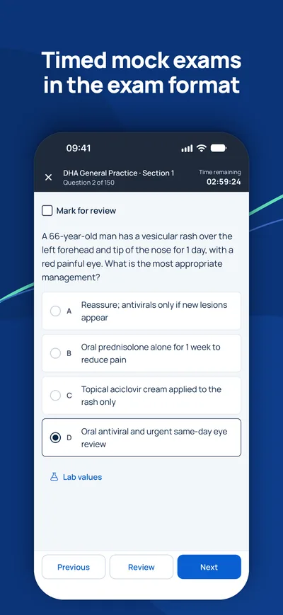
Arbeiten Sie die Kapitel Thema für Thema durch, wiederholen Sie die Lernnotizen und üben Sie dann mit zeitgesteuerten Probeprüfungen im Format Ihrer Behörde.
Fragen
Wie lange habe ich Zugang?+
Der Zugang gilt 90 Tage ab Zahlung für das gewählte Land und den gewählten Beruf.
Welche Geräte kann ich nutzen?+
Jeden Browser auf Computer, Tablet oder Handy sowie die MediFlow-App mit demselben Konto.
Kann ich das Geld zurückbekommen?+
Bitte lesen Sie vor dem Kauf unsere Erstattungsrichtlinie. Erstattungsrichtlinie

iPhone · iPad
GCC Medical Exam Prep
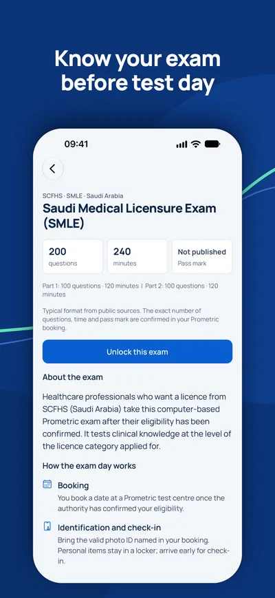
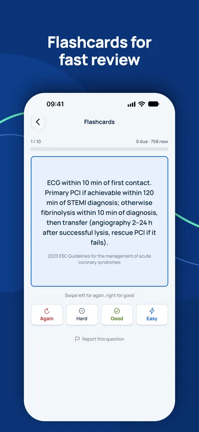
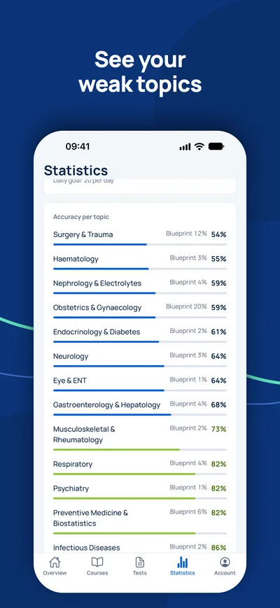
Bereiten Sie sich auf Ihrem iPhone auf DHA, DOH, MOHAP, SCFHS und weitere Zulassungsprüfungen am Golf vor.

Ist die Prüfung Ihr einziger offener Schritt?
Prüfen Sie zuerst, ob Sie die Prüfung überhaupt brauchen und welchen Titel Sie erwarten können. Ein Berater beantwortet Ihre Fragen kostenlos.
Eignungs-Check- 1Kostenlose Beratung per Video, Telefon oder WhatsApp
- 2Eignungs-Check gegen die offiziellen Anforderungen
- 3Ihr Plan mit Festpreisen und klaren nächsten Schritten