
🇧🇭 NHRA
General Medicine & GP Bahrain
General practitioners and physicians preparing for DHA, DOH, MOHAP, SMLE and other GCC licensing exams

Voorbeeldvraag
Which drug has been shown to reduce mortality in heart failure with reduced ejection fraction?
Keys: 1–5 or A–E to choose, Enter to check and continue, F to mark.
Original MediFlow content, not official exam questions. Medical review in progress.
Syllabus and high-yield notes
1. Cardiology · 4 % gewicht in het examen · 4 lessons+
- Record a 12-lead ECG within 10 minutes of first contact for chest pain. ST elevation in II, III and aVF means an inferior infarct (usually the right coronary artery): add V4R, and if the right ventricle is involved avoid nitrates and give fluids for hypotension. ST depression in V1–V3 with tall R waves may be a posterior STEMI – record V7–V9.
- STEMI: primary PCI if it can be performed within 120 minutes of diagnosis; otherwise fibrinolysis within 10 minutes of diagnosis and transfer to a PCI centre. NSTE-ACS with shock, refractory pain, acute heart failure or life-threatening arrhythmia needs angiography within 2 hours; a confirmed NSTEMI without these features within 24 hours.
- Heart failure with reduced ejection fraction is treated with four drug classes started early and up-titrated: ACE inhibitor or sacubitril/valsartan, an evidence-based beta-blocker, a mineralocorticoid receptor antagonist and an SGLT2 inhibitor. SGLT2 inhibitors also reduce heart-failure hospitalisation when the ejection fraction is mildly reduced or preserved; loop diuretics treat congestion only.
- Atrial fibrillation: haemodynamic instability calls for synchronised DC cardioversion. Stroke risk is scored with CHA₂DS₂-VA (ESC 2024; female sex no longer counts); anticoagulate at a score of 2 or more and consider it at 1. DOACs are preferred, except with a mechanical valve or moderate-to-severe mitral stenosis, where warfarin is used.
- Regular narrow-complex tachycardia in a stable patient: vagal manoeuvre first, then adenosine 6 mg as a rapid IV bolus (12 mg if needed). In pre-excited AF (WPW) avoid AV-nodal blockers – adenosine, verapamil, diltiazem, digoxin, beta-blockers and IV amiodarone; use procainamide or ibutilide, or cardioversion if unstable. Torsades de pointes responds to IV magnesium.
- Mobitz II, high-degree and complete AV block without a reversible cause need a permanent pacemaker regardless of symptoms. Syncope during exertion or while lying down, without warning, with palpitations, structural heart disease or an abnormal ECG suggests a cardiac cause and needs prompt assessment.
- Hypertension (ESC 2024): confirm with home or ambulatory readings, start most patients on a low-dose two-drug single-pill combination (RAS blocker plus calcium-channel blocker or thiazide-like diuretic) and aim for a systolic pressure of 120–129 mmHg if tolerated. The 2025 AHA/ACC guideline sets a target below 130/80 mmHg.
- Severe symptomatic aortic stenosis (peak velocity ≥ 4 m/s, mean gradient ≥ 40 mmHg, area ≤ 1 cm²) needs valve intervention; the Heart Team favours TAVI in older or high-surgical-risk patients. Endocarditis prophylaxis is limited to high-risk patients (prosthetic valve, previous endocarditis, certain congenital disease) having dental procedures that manipulate the gingiva: amoxicillin 2 g 30–60 minutes before.
- Acute pericarditis: pleuritic pain relieved by sitting forward, friction rub, widespread concave ST elevation with PR depression. Treat with an NSAID or aspirin plus colchicine for about 3 months. Hypotension, raised JVP and muffled heart sounds (Beck triad) with pulsus paradoxus mean tamponade – urgent pericardiocentesis.
- Lipids (ESC/EAS): very-high-risk patients, including everyone after an acute coronary syndrome, need LDL cholesterol below 1.4 mmol/L (55 mg/dL) and a reduction of at least 50 %. Use a high-intensity statin, add ezetimibe if the target is missed, then a PCSK9 inhibitor; measure lipoprotein(a) once in adulthood.
- Acute coronary syndromes: recognising and treating chest pain – An acute coronary syndrome (ACS) happens when an atherosclerotic plaque in a coronary artery ruptures or erodes and a thrombus forms on top of it. The thrombus narrows or blocks the artery, and the heart muscle downstream becomes ischaemic. ACS is a spectrum: unstable angina, non-ST-elevation myocardial infarction (NSTEMI) and ST-elevation myocardial infarction (STEMI). What separates them is the ECG and the troponin, not the severity of the pain.
- Chronic coronary syndromes, lipids and hypertension – Most coronary disease is chronic. Stable angina is chest discomfort brought on by exertion or emotion and relieved within a few minutes by rest or nitrate. It occurs when a fixed narrowing limits flow during increased demand. The 2024 ESC guideline calls this a chronic coronary syndrome (CCS) and stresses two parallel goals: relieve symptoms and prevent events.
- Heart failure, valve disease, pericardium and the aorta – Heart failure is a clinical syndrome: symptoms such as breathlessness, fatigue and ankle swelling, with signs such as raised jugular venous pressure and crackles, caused by a structural or functional heart abnormality that leads to raised filling pressures or low output. It is classified by left ventricular ejection fraction (LVEF), because treatment depends on it.
- Arrhythmias: atrial fibrillation, tachycardias and bradycardias – For any arrhythmia, the first question is whether the patient is stable. Adverse features in the Resuscitation Council and ERC algorithms are shock (hypotension, pallor, sweating, confusion), syncope, myocardial ischaemia and severe heart failure. An unstable tachycardia needs synchronised DC cardioversion under sedation or anaesthesia, regardless of the exact rhythm. A stable patient allows time to read the ECG carefully.
2. Respiratory · 4 % gewicht in het examen · 4 lessons+
- Asthma (GINA): SABA-only treatment is no longer recommended for adults and adolescents because it raises the risk of severe attacks. The preferred Track 1 approach uses low-dose ICS–formoterol as the reliever at every step: as needed alone at steps 1–2, and as maintenance-and-reliever therapy (MART) from step 3.
- Acute asthma: peak flow 33–50 % of best, inability to complete sentences, respiratory rate ≥ 25/min or heart rate ≥ 110/min mean a severe attack. Peak flow below 33 %, SpO₂ below 92 %, a silent chest, exhaustion, altered consciousness or a normal PaCO₂ mean a life-threatening attack; a raised PaCO₂ means near-fatal asthma.
- COPD (GOLD 2026): a post-bronchodilator FEV₁/FVC below 0.7 is required for diagnosis. Group E now means one or more moderate or severe exacerbations in the previous year. Initial therapy is a bronchodilator for group A and LABA + LAMA for groups B and E; add ICS in group E when blood eosinophils are 300/µL or more.
- COPD exacerbation: short-acting bronchodilators, prednisolone for 5 days and antibiotics for 5 days if sputum is purulent. In patients at risk of hypercapnia, aim for SpO₂ 88–92 % with controlled oxygen. NIV is indicated when pH stays below 7.35 with PaCO₂ above 6.5 kPa (49 mmHg) despite optimal treatment.
- Community-acquired pneumonia: CURB-65 guides site of care (0–1 home, 2 consider admission, 3–5 severe). A healthy outpatient receives oral amoxicillin for 5 days. Think of Legionella with hotel stays or air-conditioning exposure, confusion, diarrhoea, hyponatraemia and raised liver enzymes, and request a urinary antigen test.
- Suspected pulmonary embolism: assess clinical probability first (Wells). If PE is unlikely, measure D-dimer, using an age-adjusted cut-off (age × 10 µg/L) above 50 years; a negative result excludes PE. If PE is likely or the D-dimer is positive, perform CTPA. High-risk PE with shock is treated with systemic thrombolysis.
- Tuberculosis (WHO): use a rapid molecular test such as Xpert MTB/RIF Ultra on sputum as the initial diagnostic test; IGRA and tuberculin skin tests cannot diagnose active disease. Drug-susceptible TB is treated with 2 months of HRZE and 4 months of HR. Latent infection is treated with a rifamycin-based regimen (e.g. 4 months of rifampicin or 3 months of weekly isoniazid–rifapentine) once active TB is excluded.
- Pleural effusion: Light's criteria classify fluid as an exudate if any one is met – pleural/serum protein ratio above 0.5, pleural/serum LDH ratio above 0.6, or pleural LDH above two-thirds of the upper limit of serum LDH. In pleural infection, frank pus, organisms on Gram stain or culture, or pH below 7.2 require a chest drain.
- Pneumothorax: tension pneumothorax is a clinical diagnosis needing immediate decompression. Secondary pneumothorax (underlying lung disease such as COPD) or significant hypoxia are high-risk features and usually need a chest drain and admission; a minimally symptomatic primary pneumothorax may be managed conservatively or with aspiration.
- Interpret blood gases stepwise: pH, primary disturbance, then expected compensation. In respiratory acidosis bicarbonate rises by about 1 mmol/L per 10 mmHg (1.3 kPa) rise in PaCO₂ when acute and 3.5–4 mmol/L when chronic. Values between the two suggest acute-on-chronic hypercapnia, typical of a COPD exacerbation.
- Asthma and COPD: diagnosis, stepwise treatment and exacerbations – Asthma and chronic obstructive pulmonary disease (COPD) both narrow the airways, but they behave differently. Asthma is variable airway inflammation: symptoms come and go, and airflow limitation reverses. COPD is persistent airflow limitation, usually caused by smoking or biomass smoke, and it does not fully reverse. Spirometry measures forced expiratory volume in 1 second (FEV1) and forced vital capacity (FVC). Their ratio below 0.70 after bronchodilator confirms persistent obstruction in COPD (GOLD).
- Pneumonia and tuberculosis – Community-acquired pneumonia (CAP) is an acute infection of the lung parenchyma in a patient who was not recently in hospital. Typical features are fever, cough, purulent sputum, pleuritic pain and new focal signs, confirmed by a new opacity on chest X-ray. Streptococcus pneumoniae is the commonest cause. Haemophilus influenzae is frequent in COPD. Atypical organisms are Mycoplasma pneumoniae (young adults, epidemics, cold agglutinins), Legionella pneumophila and Chlamydophila species. Staphylococcus aureus causes severe, cavitating pneumonia after influenza.
- Pulmonary embolism, pleural disease and pneumothorax – Pulmonary embolism (PE) is usually a thrombus from the deep veins of the legs that lodges in the pulmonary arteries. Risk factors follow Virchow's triad: stasis (immobility, long travel), endothelial injury (surgery, trauma) and hypercoagulability (cancer, pregnancy, oestrogen-containing contraceptives or HRT, thrombophilia). Symptoms are breathlessness, pleuritic pain, haemoptysis, syncope; signs include tachycardia, tachypnoea and hypoxia.
- Lung cancer, interstitial lung disease, sarcoidosis, sleep apnoea and blood gases – Lung cancer is the leading cause of cancer death worldwide. About 85 % are non-small-cell lung cancer (NSCLC: adenocarcinoma, the commonest type, also in never-smokers; and squamous cell carcinoma). About 15 % are small-cell lung cancer (SCLC), strongly linked to smoking, central, fast-growing and often already spread at diagnosis. Symptoms include persistent cough, haemoptysis, weight loss, chest pain, hoarseness (recurrent laryngeal nerve), and finger clubbing.
3. Endocrinology & Diabetes · 2 % gewicht in het examen · 4 lessons+
- Diabetes is diagnosed by fasting plasma glucose ≥ 7.0 mmol/L (126 mg/dL), 2-hour OGTT glucose ≥ 11.1 mmol/L (200 mg/dL), HbA1c ≥ 6.5 % (48 mmol/mol), or random glucose ≥ 11.1 mmol/L with classic symptoms. Without symptoms, two abnormal results are needed – from the same sample or on two occasions; if two tests disagree, repeat the one above the threshold.
- Type 2 diabetes treatment (ADA 2026) is chosen by comorbidity, not HbA1c alone: an SGLT2 inhibitor for heart failure or CKD, and a GLP-1 receptor agonist or SGLT2 inhibitor with proven benefit for atherosclerotic disease. With obesity, prefer agents with high weight-loss efficacy (semaglutide, tirzepatide). Avoid pioglitazone and saxagliptin in heart failure.
- Obesity and type 2 diabetes are very common in the Gulf, and Asian and Gulf populations develop metabolic risk at lower BMI and greater waist size. Treat obesity as a chronic disease: lifestyle support, then GLP-1-based pharmacotherapy, and metabolic surgery in suitable patients who do not reach durable weight loss.
- Ramadan (IDF-DAR): stratify risk before fasting, prefer gliclazide or DPP-4 inhibitors over glibenclamide, and reduce sulfonylurea or insulin doses. Patients must break the fast at once if glucose is below 3.9 mmol/L (70 mg/dL) or above 16.7 mmol/L (300 mg/dL), or if they develop symptoms of hypoglycaemia or acute illness.
- DKA (ADA 2024 consensus): glucose ≥ 11.1 mmol/L (200 mg/dL) or known diabetes, β-hydroxybutyrate ≥ 3.0 mmol/L, and pH < 7.3 and/or bicarbonate < 18 mmol/L; SGLT2 inhibitors can cause euglycaemic DKA. Give IV fluids first; delay insulin if potassium is below 3.5 mmol/L. HHS (glucose ≥ 33.3 mmol/L, effective osmolality > 300 mOsm/kg, little ketosis) is treated mainly with 0.9 % saline.
- Hypoglycaemia is below 3.9 mmol/L (70 mg/dL); below 3.0 mmol/L (54 mg/dL) is clinically significant. A conscious patient takes 15–20 g of fast-acting glucose and rechecks after 15 minutes; an unconscious patient receives glucagon or IV glucose. Sulfonylurea-induced hypoglycaemia can recur for many hours and needs observation.
- Thyroid: subclinical hypothyroidism with TSH below 10 mU/L is usually rechecked rather than treated. Use propylthiouracil in the first trimester and carbimazole or methimazole otherwise. In thyroid storm give a beta-blocker, a thionamide, then iodine at least 1 hour later, plus hydrocortisone. Thyroid nodules: check TSH and ultrasound; FNA for suspicious nodules ≥ 1 cm.
- Adrenal: suspected adrenal crisis (hypotension, hyponatraemia, hyperkalaemia, hypoglycaemia) needs hydrocortisone 100 mg IV and saline immediately – never wait for test results. Screen for Cushing syndrome with the 1 mg overnight dexamethasone test, late-night salivary cortisol or 24-hour urinary free cortisol; for primary aldosteronism with the aldosterone-to-renin ratio; for phaeochromocytoma with plasma or urinary metanephrines, followed by alpha-blockade before surgery.
- Calcium and bone: primary hyperparathyroidism shows high calcium with high or inappropriately normal PTH; a calcium-to-creatinine clearance ratio below 0.01 points to familial hypocalciuric hypercalcaemia, which needs no surgery. Vitamin D deficiency is very common in the Gulf despite sunshine. Osteoporosis (T-score ≤ −2.5) is usually treated first with an oral bisphosphonate.
- Pituitary and PCOS: prolactinomas, even macroadenomas, are treated first with cabergoline; exclude pregnancy, hypothyroidism and drugs as causes of raised prolactin. PCOS is diagnosed by two of three Rotterdam criteria after excluding mimics; letrozole is first-line for anovulatory infertility.
- Diabetes mellitus: diagnosis and long-term treatment – Diabetes mellitus is chronic hyperglycaemia caused by too little insulin, too little insulin effect, or both. Type 1 diabetes is autoimmune destruction of pancreatic beta cells; patients need insulin from diagnosis and are prone to ketoacidosis. Type 2 diabetes combines insulin resistance (linked to obesity, inactivity and genes) with gradually failing beta cells. Other types include gestational diabetes, monogenic diabetes (MODY), and diabetes from pancreatic disease or drugs such as steroids.
- Diabetic emergencies: DKA, HHS and hypoglycaemia – Diabetic ketoacidosis (DKA) arises when insulin is absent or very low. Without insulin, cells cannot use glucose, so the body breaks down fat; the liver turns fatty acids into ketone bodies, which are acids. Glucose rises, causing an osmotic diuresis and severe dehydration. DKA is typical of type 1 diabetes, often triggered by infection, missed insulin, or new diagnosis, but it also occurs in type 2 and with SGLT2 inhibitors, where glucose may be near normal (euglycaemic DKA).
- Thyroid disease: hypothyroidism, thyrotoxicosis, nodules and pregnancy – The hypothalamus releases TRH, which stimulates pituitary TSH, which stimulates the thyroid to make thyroxine (T4) and some triiodothyronine (T3). T4 and T3 feed back to suppress TSH. Because TSH responds in a log-linear way to small changes in free T4, TSH is the best single screening test in primary thyroid disease. The exceptions are pituitary disease and recent treatment changes, where free T4 is more reliable.
- Adrenal, pituitary, calcium and bone, and reproductive endocrinology – The adrenal cortex makes cortisol (stress hormone, regulated by ACTH), aldosterone (salt and potassium balance, regulated by renin–angiotensin) and adrenal androgens. The medulla makes adrenaline and noradrenaline. Most adrenal disorders are disorders of excess or deficiency of one of these.
4. Gastroenterology & Hepatology · 4 % gewicht in het examen · 4 lessons+
- Typical reflux without alarm features is treated empirically: lifestyle advice plus a once-daily PPI for 8 weeks. Dysphagia, unintentional weight loss, GI bleeding, iron deficiency anaemia, persistent vomiting, or age 55 or over with weight loss and dyspepsia call for urgent upper GI endoscopy instead.
- Clarithromycin resistance in the Gulf region is high, so first-line H. pylori eradication is 14-day bismuth quadruple therapy (PPI, bismuth, tetracycline, metronidazole). Stop PPIs 2 weeks and antibiotics or bismuth 4 weeks before a urea breath test or stool antigen test, and confirm eradication in every treated patient.
- In upper GI bleeding, a Glasgow-Blatchford score of 0–1 allows outpatient management. Otherwise resuscitate, transfuse restrictively (Hb threshold about 70 g/L, 80 g/L with cardiovascular disease) and perform endoscopy within 24 hours; suspected variceal bleeding needs terlipressin and antibiotics before endoscopy.
- Acute severe ulcerative colitis (Truelove and Witts: 6 or more bloody stools a day plus tachycardia, fever, anaemia or raised ESR/CRP) needs admission, IV corticosteroids, LMWH thromboprophylaxis, stool tests including C. difficile, and no opioids, antidiarrhoeals or NSAIDs. Failure to respond by day 3 triggers rescue therapy (infliximab or ciclosporin) or colectomy.
- Coeliac serology (tTG-IgA plus total IgA) is valid only while the patient eats gluten. If total IgA is low, use IgG-based tests (DGP-IgG or tTG-IgG); in adults, positive serology is confirmed by duodenal biopsy before a lifelong gluten-free diet is started.
- Hepatitis serology: HBsAg = current infection; IgM anti-HBc = acute infection and the only marker in the window period; anti-HBs alone = vaccination; anti-HBc plus anti-HBs = resolved infection. A reactive anti-HCV shows exposure only, and HCV RNA confirms current infection. Hepatitis E can cause fulminant liver failure in pregnancy; hepatitis D occurs only with HBsAg.
- Cirrhosis complications: ascitic neutrophils of 250/mm³ or more = SBP (cefotaxime or ceftriaxone plus IV albumin); overt hepatic encephalopathy = lactulose, adding rifaximin after a recurrence; clinically significant portal hypertension = non-selective beta-blocker, preferably carvedilol; HCC surveillance with ultrasound, with or without AFP, every 6 months.
- Acute pancreatitis is diagnosed with 2 of 3: typical pain, lipase or amylase at least 3 times the upper limit of normal, characteristic imaging. Give moderate, goal-directed fluids (Ringer's lactate) rather than aggressive volumes, feed orally early, avoid prophylactic antibiotics, reserve urgent ERCP for cholangitis, and remove the gallbladder during the same admission after mild gallstone pancreatitis.
- Colorectal cancer screening at average risk runs from 45 to 75 years (FIT every year or colonoscopy every 10 years, USPSTF 2021). One first-degree relative diagnosed before 60 means colonoscopy every 5 years from age 40, or 10 years before the relative's age at diagnosis if earlier; Lynch syndrome needs colonoscopy every 1–2 years from age 20–25.
- Read liver tests as a pattern: ALT-predominant rise = hepatocellular; ALP with raised GGT = cholestatic or biliary; ALP with normal GGT = bone; AST:ALT above 2 suggests alcohol; isolated unconjugated hyperbilirubinaemia with a normal blood count and reticulocytes = Gilbert syndrome.
- Upper GI tract: reflux, dyspepsia, Helicobacter pylori and upper GI bleeding – Most upper gastrointestinal complaints in general practice are benign: heartburn, acid regurgitation and epigastric discomfort after meals. The task of the doctor is twofold. First, decide whether the patient can be treated empirically, or whether an alarm feature means cancer or a complication must be excluded by endoscopy. Second, find and remove treatable causes, mainly Helicobacter pylori and non-steroidal anti-inflammatory drugs (NSAIDs). This chapter follows that logic and ends with the emergency of upper GI bleeding.
- Lower GI tract: inflammatory bowel disease, IBS, coeliac disease and colorectal cancer screening – Chronic diarrhoea, abdominal pain and rectal bleeding are common reasons for referral. The central question is whether the cause is organic (inflammation, malabsorption, neoplasia) or functional. Simple tests separate them well: full blood count, CRP, coeliac serology, faecal calprotectin, and colonoscopy when indicated. This chapter covers the four diseases examiners test most: ulcerative colitis, Crohn's disease, irritable bowel syndrome and coeliac disease, and then colorectal cancer screening.
- The liver: reading liver tests, viral hepatitis and inherited or autoimmune liver disease – Liver blood tests are among the most frequently ordered tests and the most frequently misread. The term 'liver function tests' is misleading, because ALT, AST and ALP measure injury, not function. True synthetic function is shown by albumin, INR (prothrombin time) and bilirubin clearance. Learn to read the pattern first, then think of the cause.
- Cirrhosis and its complications – Cirrhosis is the end stage of chronic liver injury: diffuse fibrosis with regenerative nodules that distort the liver architecture. The leading causes are alcohol, metabolic dysfunction-associated steatotic liver disease (MASLD, formerly NAFLD), and chronic hepatitis B and C. Cirrhosis can be compensated for years. Decompensation means the appearance of ascites, variceal bleeding, hepatic encephalopathy or jaundice, and it sharply reduces survival.
5. Nephrology & Electrolytes · 4 % gewicht in het examen · 5 lessons+
- KDIGO defines AKI as a creatinine rise of ≥ 26.5 µmol/L (0.3 mg/dL) within 48 hours, a rise to ≥ 1.5 times baseline within 7 days, or urine output below 0.5 mL/kg/h for 6 hours. Stage by the worse of the creatinine or urine-output criteria: stage 2 is 2.0–2.9 times baseline, stage 3 is 3 times or more, creatinine ≥ 353.6 µmol/L (4.0 mg/dL), or dialysis.
- In AKI, look for volume depletion, nephrotoxins and obstruction. Stop NSAIDs and withhold ACE inhibitors, ARBs, diuretics and metformin during acute illness ('sick-day rules'). FENa below 1 % suggests a prerenal cause. Urgent dialysis is needed for refractory hyperkalaemia, acidosis or fluid overload, uraemic pericarditis or encephalopathy, and dialysable poisons.
- CKD is classified by cause, GFR category (G1–G5) and albuminuria (A1–A3). KDIGO 2024 core therapy: an ACE inhibitor or ARB at the maximum tolerated dose when albuminuria is present, an SGLT2 inhibitor when eGFR is 20 or more, and a statin for most adults. A creatinine rise of up to 30 % after starting a RAS blocker is expected and not a reason to stop. Diabetes is the leading cause of CKD in the Gulf.
- Nephrotic syndrome: proteinuria above 3.5 g/day, albumin below 30 g/L, oedema and hyperlipidaemia, with a high risk of thrombosis and infection. Minimal change disease dominates in children; membranous nephropathy (anti-PLA2R antibodies) and FSGS in adults. Nephritic syndrome: haematuria with red-cell casts, hypertension, oliguria and modest proteinuria.
- Haematuria after a sore throat: within 1–3 days with normal complement suggests IgA nephropathy; after 1–3 weeks with low C3 suggests post-streptococcal glomerulonephritis. Rapidly rising creatinine with haemoptysis is a pulmonary–renal syndrome: test ANCA and anti-GBM antibodies urgently.
- Urinary tract infection: uncomplicated cystitis in women is treated with nitrofurantoin for 5 days, single-dose fosfomycin or pivmecillinam; avoid fluoroquinolones. Asymptomatic bacteriuria is treated only in pregnancy and before procedures that breach the urinary-tract mucosa. Fever with an obstructed kidney is a urological emergency.
- Kidney stones are common in the hot Gulf climate. Use low-dose non-contrast CT for diagnosis and NSAIDs as first-line analgesia. Distal ureteric stones over 5 mm may pass with tamsulosin (medical expulsive therapy). An infected obstructed kidney needs urgent decompression by stent or nephrostomy. Prevent recurrence with enough fluid for at least 2–2.5 L of urine a day.
- Hyponatraemia with severe symptoms (seizures, coma) is treated with a 3 % saline bolus (e.g. 150 mL over 20 minutes), repeated until sodium rises by about 5 mmol/L. In chronic hyponatraemia, limit correction to 10 mmol/L in 24 hours, or 8 mmol/L in high-risk patients (alcohol use, malnutrition, hypokalaemia, liver disease), to prevent osmotic demyelination. Correct chronic hypernatraemia slowly with hypotonic fluid.
- Potassium: hyperkalaemia with ECG changes needs IV calcium first, then insulin–glucose and nebulised salbutamol, then potassium removal. Exclude pseudohyperkalaemia (haemolysis, very high platelet or white-cell counts, delayed processing) when the result does not fit the clinical picture. Hypokalaemia that does not respond to replacement usually reflects low magnesium.
- Acid–base: calculate the anion gap (Na⁺ − Cl⁻ − HCO₃⁻, normal about 8–12 mmol/L). In normal-gap acidosis, a negative urine anion gap points to gut bicarbonate loss and a positive one to renal tubular acidosis. Vomiting causes chloride-responsive metabolic alkalosis (urine chloride below 20 mmol/L), treated with saline and potassium. Adjust drug doses to kidney function: stop metformin below eGFR 30 and avoid nitrofurantoin below 45.
- Acute kidney injury: definition, causes and management – Acute kidney injury (AKI) is a sudden fall in kidney function, measured by a rise in serum creatinine or a fall in urine output. It is common in hospital, especially with sepsis, dehydration, surgery and nephrotoxic drugs, and it increases mortality even when mild. Creatinine lags behind the injury by a day or two, so a 'normal' creatinine early in an illness does not exclude AKI.
- Chronic kidney disease and glomerular disease – Chronic kidney disease (CKD) is an abnormality of kidney structure or function present for more than 3 months with implications for health. In practice, that means an eGFR below 60 mL/min/1.73 m2 or markers of damage such as albuminuria, for at least 3 months. KDIGO stages CKD by cause, GFR (G1–G5) and albuminuria (A1–A3). The two together predict progression and cardiovascular risk better than either alone.
- Urinary tract infection and kidney stones – Urinary tract infection (UTI) is one of the commonest infections. Escherichia coli causes about 75–80 % of community cases; others are Klebsiella, Proteus (alkaline urine, struvite stones) and Staphylococcus saprophyticus in young women. Infection is classified by site (cystitis in the bladder, pyelonephritis in the kidney) and as uncomplicated (healthy non-pregnant women with a normal urinary tract) or complicated (men, pregnancy, catheters, obstruction, structural abnormality, immunosuppression).
- Sodium, potassium and other electrolytes – Sodium disorders are really water disorders. Serum sodium reflects the balance of water relative to sodium. The brain is most at risk: low sodium makes cells swell, high sodium makes them shrink. Therefore speed matters as much as size, both in how fast the abnormality developed and in how fast it is corrected.
- Acid–base disorders and safe prescribing in kidney disease – Acid–base analysis answers three questions: is the blood acidaemic or alkalaemic, is the main problem respiratory or metabolic, and is the body compensating as expected? Normal values: pH 7.35–7.45, PaCO2 4.7–6.0 kPa (35–45 mmHg), bicarbonate 22–26 mmol/L.
6. Haematology · 3 % gewicht in het examen · 4 lessons+
- Start every microcytic anaemia with ferritin: below 15 µg/L confirms iron deficiency, values up to about 45 µg/L make it likely, and inflammation raises ferritin. Microcytosis with a normal ferritin and a high red cell count (Mentzer index below 13) suggests thalassaemia trait; check HbA2 by HPLC or electrophoresis, a key part of premarital screening in the Gulf.
- Iron deficiency anaemia always needs a cause: coeliac serology for everyone and bidirectional endoscopy for men and postmenopausal women. Treat with one oral iron tablet daily or on alternate days (better absorbed and tolerated than several doses a day), use IV iron if oral iron fails or is not tolerated, and continue for about 3 months after the Hb normalises.
- B12 deficiency with neurological signs is treated with hydroxocobalamin 1 mg IM on alternate days until there is no further improvement, then every 2 months. When B12 and folate are both low, start B12 first, because folic acid alone can precipitate subacute combined degeneration of the cord; anti-intrinsic factor antibodies confirm pernicious anaemia.
- Haemolysis = anaemia with raised reticulocytes, unconjugated bilirubin and LDH, and low haptoglobin. A positive DAT means immune haemolysis (warm AIHA: spherocytes, prednisolone 1 mg/kg). A negative DAT with bite or blister cells after fava beans, primaquine, dapsone, rasburicase or nitrofurantoin points to G6PD deficiency; retest G6PD about 3 months after an acute episode.
- Transfusion-dependent beta-thalassaemia: transfuse to keep the pre-transfusion Hb at 95–105 g/L and start chelation after 10–20 transfusions or when ferritin exceeds 1,000 µg/L. Deferiprone can cause agranulocytosis (weekly neutrophil count), deferasirox renal and hepatic toxicity, and deferoxamine ocular and auditory toxicity.
- Sickle cell disease: treat painful crises with rapid opioid analgesia and fluids. Fever, chest pain, hypoxia and a new infiltrate mean acute chest syndrome (oxygen, antibiotics including a macrolide, incentive spirometry, transfusion, with exchange transfusion if severe or deteriorating). A sudden Hb fall with low reticulocytes suggests parvovirus B19 aplastic crisis; with a rapidly enlarging spleen, splenic sequestration. Hydroxyurea, penicillin prophylaxis and vaccination reduce complications.
- The bleeding pattern guides diagnosis: mucocutaneous bleeding (epistaxis, menorrhagia, bruising) suggests a platelet or von Willebrand factor problem; haemarthroses and muscle haematomas suggest a coagulation factor deficiency (haemophilia: prolonged APTT, normal PT). In adult ITP, treat platelets below 30 × 10⁹/L or bleeding with corticosteroids; TTP needs urgent plasma exchange, not platelets; suspected HIT means stopping all heparin and starting a non-heparin anticoagulant.
- Warfarin: a high INR without bleeding is managed by withholding doses, adding low-dose oral vitamin K if the INR is above 8 or bleeding risk is high; major bleeding needs prothrombin complex concentrate plus IV vitamin K 5–10 mg. Dabigatran is reversed by idarucizumab and factor Xa inhibitors by andexanet alfa or PCC. DOACs are contraindicated with mechanical heart valves and moderate-to-severe mitral stenosis, and warfarin is preferred in triple-positive antiphospholipid syndrome.
- Venous thromboembolism: use the Wells score; if DVT or PE is unlikely, a normal D-dimer excludes it, and if likely, image directly. PE with sustained hypotension is high-risk and needs systemic thrombolysis. Treat provoked VTE for 3 months and consider extended treatment after unprovoked VTE; use a DOAC or LMWH in cancer and LMWH in pregnancy.
- Haematological malignancy clues: smudge cells in an older adult = CLL (often watch and wait); Philadelphia chromosome t(9;22) = CML (imatinib); t(15;17) with DIC = acute promyelocytic leukaemia (all-trans retinoic acid at once); Reed–Sternberg cells = Hodgkin lymphoma. In any transfusion reaction stop the transfusion first; breathlessness with hypertension and fluid overload suggests TACO, with hypotension and fever TRALI.
- Approach to anaemia: iron deficiency, thalassaemia trait and macrocytic anaemia – Anaemia is a reduced haemoglobin concentration: below about 130 g/L in men and 120 g/L in non-pregnant women (WHO). It is a sign, not a diagnosis, so the task is always to find the cause. The most useful first step is the mean cell volume (MCV), which divides anaemia into microcytic (MCV below about 80 fL), normocytic and macrocytic (above about 100 fL). The reticulocyte count then tells you whether the marrow is responding (high in bleeding and haemolysis) or failing (low in deficiency and marrow disease).
- Haemolysis, thalassaemia major, sickle cell disease and G6PD deficiency – Haemolysis means premature destruction of red cells. The laboratory pattern is the same whatever the cause: anaemia, raised reticulocytes (the marrow compensates), raised unconjugated bilirubin and lactate dehydrogenase (LDH) from broken cells, and low haptoglobin, which binds free haemoglobin and is consumed. Intravascular haemolysis adds haemoglobinuria and haemosiderinuria. The direct antiglobulin test (DAT, Coombs test) then splits causes into immune and non-immune.
- Bleeding disorders, thrombocytopenia and anticoagulation – Haemostasis has two stages. Primary haemostasis is the formation of a platelet plug, which needs normal platelets and von Willebrand factor (VWF). Secondary haemostasis is the coagulation cascade, which converts fibrinogen into fibrin to stabilise the plug. The bleeding pattern tells you which stage is defective. Mucocutaneous bleeding, such as epistaxis, gum bleeding, menorrhagia, petechiae and easy bruising, suggests a platelet or VWF problem. Deep bleeding into joints (haemarthrosis) and muscles, or delayed bleeding after surgery, suggests a coagulation factor deficiency.
- Haematological malignancy and transfusion medicine – Haematological cancers often first appear as an abnormal blood count. A generalist does not need to know chemotherapy protocols, but must recognise the patterns, know which findings are emergencies, and refer appropriately. This chapter covers leukaemias, lymphomas, myeloma, oncological emergencies and safe transfusion.
7. Infectious Diseases · 2 % gewicht in het examen · 4 lessons+
- Sepsis (Surviving Sepsis Campaign 2026): screen ward patients with NEWS2, MEWS or SIRS rather than qSOFA alone. Give antimicrobials immediately, ideally within 1 hour, for septic shock or probable sepsis, and within 3 hours after rapid assessment when sepsis is only possible. For hypoperfusion give at least 30 mL/kg balanced crystalloid in the first 3 hours, then norepinephrine (it may be started peripherally) to a MAP of 65 mmHg, or 60–65 mmHg from age 65.
- Suspected bacterial meningitis: take blood cultures and give dexamethasone with the first dose of ceftriaxone or cefotaxime without delay; add ampicillin or amoxicillin for Listeria in patients over 50 or immunocompromised, and vancomycin where resistant pneumococci occur. CT before lumbar puncture is needed only with focal signs, new seizures, markedly reduced consciousness, papilloedema or immunocompromise, and antibiotics are never delayed for imaging.
- HIV: screen with a fourth-generation antigen/antibody test; if it is reactive and the differentiation assay is negative, an HIV-1 RNA test detects acute infection. PEP must start within 72 hours (ideally within 24), lasts 28 days and uses an integrase-inhibitor regimen such as bictegravir/emtricitabine/tenofovir alafenamide; it is not needed when the source has a sustained undetectable viral load.
- STIs (CDC 2021): gonorrhoea = ceftriaxone 500 mg IM once (1 g from 150 kg), plus doxycycline if chlamydia is not excluded; chlamydia = doxycycline 100 mg twice daily for 7 days, or azithromycin 1 g once in pregnancy; early syphilis = benzathine penicillin G 2.4 million units IM once. Fever and myalgia within 24 hours of syphilis treatment is a Jarisch–Herxheimer reaction, not penicillin allergy.
- Fever after travel to a malaria area is malaria until proven otherwise: repeat thick and thin films or rapid diagnostic tests. Severe falciparum malaria (impaired consciousness, renal failure, jaundice, high parasitaemia) needs IV artesunate followed by a full oral artemisinin-based course; P. vivax and P. ovale also need primaquine or tafenoquine against liver hypnozoites, but only after G6PD testing.
- Brucellosis is endemic in the Gulf and is acquired from raw milk or contact with camels, goats and sheep: undulant fever, sweats, arthralgia, sacroiliitis and hepatosplenomegaly. Diagnose with prolonged blood cultures (warn the laboratory) and serology; treat with doxycycline for 6 weeks plus streptomycin or gentamicin, or plus rifampicin; spondylitis needs at least 12 weeks.
- MERS-CoV: consider it in fever with respiratory illness within 14 days of contact with dromedary camels, raw camel products or a confirmed case, especially in healthcare workers. Isolate early, confirm with real-time RT-PCR, preferably on a lower respiratory tract specimen, and give supportive care; there is no licensed vaccine or specific antiviral.
- Typhoid: gradually rising fever, headache, abdominal pain, relative bradycardia and rose spots after travel to South Asia. Blood culture is the first test. Infections acquired in Pakistan are often extensively drug-resistant (resistant to fluoroquinolones and ceftriaxone), so uncomplicated cases receive azithromycin and complicated cases a carbapenem.
- Skin and bone: pain out of proportion to the skin findings, rapid spread, crepitus or systemic toxicity suggests necrotising fasciitis and needs immediate surgical debridement. Osteomyelitis is mostly caused by Staphylococcus aureus, but Salmonella is characteristic in sickle cell disease; MRI is the most sensitive imaging test, and a positive probe-to-bone test supports the diagnosis in diabetic foot ulcers.
- Antibiotic stewardship: do not treat asymptomatic bacteriuria except in pregnancy or before urological procedures; prefer WHO AWaRe Access antibiotics, review and de-escalate at 48–72 hours, and treat C. difficile with fidaxomicin or oral vancomycin. For Hajj and Umrah, Saudi Arabia requires quadrivalent meningococcal (ACWY) vaccination at least 10 days before arrival.
- Sepsis, septic shock and antibiotic stewardship – Sepsis kills more hospital patients than most single diseases, and the main lever for survival is early recognition. The Sepsis-3 definition (2016) describes sepsis as life-threatening organ dysfunction caused by a dysregulated host response to infection. Organ dysfunction is operationalised as an acute rise in the SOFA (Sequential Organ Failure Assessment) score of 2 points or more. Septic shock is the subset of sepsis with circulatory and metabolic failure: vasopressors are required to keep the mean arterial pressure at 65 mmHg or more, and serum lactate exceeds 2 mmol/L despite adequate fluid resuscitation.
- Central nervous system, skin, soft tissue and bone infections – Bacterial meningitis is an emergency in which every hour of delay worsens outcome. The classic triad is fever, neck stiffness and altered mental status, but fewer than half of patients have all three; nearly all have at least two of headache, fever, neck stiffness and altered consciousness. A non-blanching petechial or purpuric rash suggests Neisseria meningitidis. In adults, the main pathogens are Streptococcus pneumoniae and N. meningitidis; Listeria monocytogenes matters in people over 50, pregnant women, alcohol misusers and the immunocompromised.
- HIV, post-exposure prophylaxis and sexually transmitted infections – HIV infection is now a manageable chronic condition when diagnosed early and treated with antiretroviral therapy (ART). Undetectable viral load means the virus is not sexually transmitted (undetectable = untransmittable). The challenge is diagnosis: primary HIV infection often presents 2–4 weeks after exposure as a glandular fever-like illness with fever, sore throat, lymphadenopathy, maculopapular rash and mouth ulcers. This is the stage when viral load and infectivity are highest.
- Travel, tropical and Gulf-region infections and vaccination – Gulf countries host millions of migrant workers and travellers, and receive millions of pilgrims each year. A doctor practising there will regularly see imported malaria and typhoid, as well as locally endemic infections such as brucellosis and MERS-CoV. The approach always starts with the exposure history: where the patient travelled or came from, when, what they ate and drank, which animals they touched, and whether they took prophylaxis or had vaccines.
8. Neurology · 3 % gewicht in het examen · 4 lessons+
- Acute ischaemic stroke (AHA/ASA 2026): check glucose and do a non-contrast CT; give IV thrombolysis within 4.5 hours for disabling deficits (tenecteplase 0.25 mg/kg or alteplase 0.9 mg/kg) once BP is below 185/110 mmHg, then keep it below 180/105 mmHg for 24 hours. Thrombolysis at 4.5–9 hours or in wake-up stroke needs imaging evidence of salvageable tissue; thrombectomy for anterior-circulation large-vessel occlusion is offered up to 24 hours with imaging selection.
- TIA: NICE advises against using ABCD2 to judge risk or urgency, because it misses high-risk causes such as atrial fibrillation, carotid stenosis and crescendo TIAs. Give aspirin 300 mg immediately (unless the patient is anticoagulated or has a bleeding disorder) and arrange specialist assessment within 24 hours. After a minor stroke or high-risk TIA, aspirin plus clopidogrel for 21 days, then a single antiplatelet, reduces early recurrence; symptomatic carotid stenosis of 50–99 % needs endarterectomy, ideally within 2 weeks.
- Headache red flags: thunderclap onset, new headache after 50, fever with neck stiffness, papilloedema, focal signs, headache worse lying down or with Valsalva, immunosuppression or cancer, and jaw claudication. Thunderclap headache needs CT; if a CT within 6 hours of onset is normal, LP is usually not needed, but after 6 hours LP is done at least 12 hours after onset to look for xanthochromia. Suspected giant cell arteritis needs prednisolone the same day.
- Migraine: treat attacks with a triptan plus an NSAID or paracetamol and avoid opioids; offer prophylaxis (propranolol, amitriptyline or topiramate) for frequent or disabling attacks. Topiramate is teratogenic and reduces hormonal contraceptive efficacy, combined hormonal contraception is contraindicated in migraine with aura, and simple analgesics on 15 or more days a month (triptans or opioids on 10 or more) cause medication overuse headache. Cluster attacks respond to 100 % oxygen and subcutaneous or nasal triptans; verapamil is used for prevention.
- Convulsive status epilepticus: at 5 minutes give a benzodiazepine (IV lorazepam, or buccal or IM midazolam) and repeat once; if seizures continue, give IV levetiracetam, fosphenytoin or valproate, and escalate to general anaesthesia for refractory status. Always check glucose. In women and girls who can become pregnant, prefer lamotrigine or levetiracetam to the highly teratogenic valproate.
- Parkinson's disease is a clinical diagnosis (bradykinesia with rest tremor or rigidity). Levodopa is first-line when motor symptoms affect quality of life, and dopamine agonists can cause impulse control disorders. In hospital, give Parkinson's medicines on time, never stop them abruptly, and avoid dopamine antagonists such as metoclopramide, haloperidol and prochlorperazine; domperidone is the antiemetic of choice.
- Neuroimmunology: MS is supported by MRI lesions disseminated in space and time and by CSF oligoclonal bands; relapses are treated with high-dose methylprednisolone. Guillain–Barré syndrome (ascending weakness, areflexia, raised CSF protein with a normal cell count) needs serial FVC and IVIG or plasma exchange, not steroids. Myasthenia gravis (fatigable ptosis and diplopia, anti-AChR antibodies) needs a CT chest for thymoma and is worsened by aminoglycosides, fluoroquinolones, macrolides, magnesium and beta-blockers.
- Dementia: look for reversible contributors (B12, folate, TSH, calcium, glucose, renal and liver function, full blood count, medication review, depression). Offer donepezil, rivastigmine or galantamine for mild-to-moderate Alzheimer's disease and memantine for severe disease. In dementia with Lewy bodies (fluctuating cognition, visual hallucinations, parkinsonism, REM sleep behaviour disorder) use donepezil or rivastigmine and avoid antipsychotics because of severe neuroleptic sensitivity.
- Painful diabetic neuropathy: offer amitriptyline, duloxetine, pregabalin or gabapentin. Foot drop with weak eversion but preserved inversion and ankle reflex points to common peroneal nerve palsy at the fibular head rather than an L5 root lesion. Back pain with leg weakness or a sensory level in a patient with cancer suggests metastatic spinal cord compression (dexamethasone 16 mg and whole-spine MRI within 24 hours); saddle anaesthesia with bladder or bowel dysfunction suggests cauda equina syndrome and needs emergency MRI.
- Raised intracranial pressure: headache worse in the morning and on lying down, vomiting, papilloedema and a falling GCS; hypertension, bradycardia and irregular breathing (Cushing triad) signal imminent herniation. Nurse head-up at 30°, give osmotherapy (mannitol or hypertonic saline), get urgent neurosurgical review and avoid lumbar puncture while a mass lesion is possible. Idiopathic intracranial hypertension (typically obese women, normal MRI and MR venography, opening pressure above 25 cmH₂O) threatens vision and is treated with weight loss and acetazolamide.
- Stroke and transient ischaemic attack – Stroke is a sudden focal neurological deficit of vascular origin. About 85 percent of strokes are ischaemic (arterial occlusion by thrombus or embolus) and about 15 percent are haemorrhagic (intracerebral or subarachnoid bleeding). Clinically the two cannot be separated reliably, so imaging is mandatory before any antithrombotic treatment. The key principle is 'time is brain': about 1.9 million neurons are lost every minute of an untreated large-vessel occlusion.
- Headache, subarachnoid haemorrhage and raised intracranial pressure – Most headaches are primary: migraine, tension-type and cluster headache. The doctor's job is to spot the few secondary headaches that are dangerous, such as subarachnoid haemorrhage, meningitis, giant cell arteritis, raised intracranial pressure and venous sinus thrombosis. A careful history with attention to red flags does most of the work.
- Epilepsy, status epilepticus, Parkinson's disease and dementia – A seizure is a transient event caused by abnormal, excessive synchronous neuronal activity. Epilepsy is an enduring tendency to unprovoked seizures: two unprovoked seizures more than 24 hours apart, or one with a high recurrence risk (for example an epileptiform EEG or a structural lesion). After a first seizure, take a careful eyewitness history, check glucose, electrolytes, calcium and ECG (to exclude cardiac syncope), and arrange specialist review, EEG and MRI. Syncope is brief, with rapid recovery and little confusion; seizures cause post-ictal confusion, lateral tongue biting and sometimes incontinence.
- Neuroimmunology, neuromuscular disease, peripheral nerves and spinal emergencies – Localisation is the first step in neurology. Ask whether the lesion is in the brain, spinal cord, nerve root, peripheral nerve, neuromuscular junction or muscle. Upper motor neurone (UMN) lesions cause spasticity, hyperreflexia, extensor plantar responses and weakness in a 'pyramidal' pattern (arm extensors and leg flexors). Lower motor neurone (LMN) lesions cause wasting, fasciculation, hypotonia and reduced or absent reflexes. Neuromuscular junction disorders cause fatigable weakness without sensory loss, and myopathies cause proximal weakness with preserved reflexes.
9. Psychiatry · 1 % gewicht in het examen · 5 lessons+
- Serotonin syndrome starts within hours of adding a serotonergic drug and shows clonus, hyperreflexia, mydriasis and diarrhoea; neuroleptic malignant syndrome develops over days after a dopamine antagonist with lead-pipe rigidity, slow reflexes and a very high CK. In both, stop the culprit drugs; benzodiazepines and cyproheptadine help serotonin syndrome, dantrolene or bromocriptine are used in severe NMS.
- Lithium has a narrow therapeutic range: aim for 0.6–0.8 mmol/L (12 hours after the dose) when starting, check levels every 3 months in the first year and renal function, thyroid function and calcium every 6 months. Dehydration, thiazides, ACE inhibitors, ARBs and NSAIDs precipitate toxicity; long-term effects include hypothyroidism, hyperparathyroidism and nephrogenic diabetes insipidus.
- Mania that appears on antidepressant monotherapy indicates bipolar disorder: stop the antidepressant and start an antipsychotic (haloperidol, olanzapine, quetiapine or risperidone). Lithium is the first-choice long-term mood stabiliser; valproate must not be started in women or girls who could become pregnant.
- Extrapyramidal effects follow a timeline: acute dystonia within hours to days (parenteral anticholinergic), akathisia within days to weeks (reduce dose, propranolol), parkinsonism within weeks and tardive dyskinesia after months to years (review or switch, VMAT2 inhibitor). Olanzapine and clozapine carry the highest metabolic risk; check weight, HbA1c or fasting glucose and lipids at baseline, 12 weeks and yearly.
- Clozapine is reserved for schizophrenia that has not responded to two antipsychotics, at least one of them second-generation. It requires regular blood counts for neutropenia and agranulocytosis; also watch for myocarditis in the first 2 months, severe constipation, hypersalivation and seizures. Tobacco smoke induces CYP1A2, so stopping smoking can raise clozapine levels by about half.
- Psychological therapy comes first in anxiety-related disorders: CBT for generalised anxiety and panic disorder (an SSRI such as sertraline if drug treatment is preferred), CBT with exposure and response prevention for OCD, and trauma-focused CBT or EMDR for PTSD. Benzodiazepines are only for short-term crises and are not used to prevent or treat PTSD.
- Alcohol withdrawal: tremor and sweating from 6–12 hours, seizures at 12–48 hours and delirium tremens at 48–72 hours after the last drink. Treat with a benzodiazepine, and give parenteral thiamine before any glucose to anyone at risk of Wernicke encephalopathy (confusion, ataxia, eye-movement abnormalities).
- Opioid intoxication causes pinpoint pupils, drowsiness and respiratory depression: give naloxone and keep observing, because its effect is shorter than that of most opioids. Opioid withdrawal causes dilated pupils, yawning, lacrimation, rhinorrhoea, piloerection, cramps and diarrhoea; it is distressing but rarely dangerous and is managed with methadone or buprenorphine.
- Delirium is acute, fluctuating and marked by inattention; dementia is gradual with attention preserved early on. Capacity is decision-specific: the person must understand, retain, use or weigh the information and communicate a choice, and neither a psychiatric diagnosis nor an unwise decision by itself means incapacity.
- Previous self-harm is the strongest single predictor of suicide. Everyone who presents after self-harm needs a psychosocial assessment by a mental health professional; risk scales and low, medium or high labels must not decide who is offered treatment.
- Depression and bipolar disorder: recognition, antidepressants and lithium – Depression is the commonest psychiatric problem a general doctor will meet. A depressive episode means low mood or loss of interest and pleasure (anhedonia) for most of the day, nearly every day, for at least two weeks, together with other symptoms: poor sleep or oversleeping, change in appetite or weight, fatigue, poor concentration, feelings of worthlessness or guilt, psychomotor slowing or agitation, and thoughts of death or suicide. ICD-11 and DSM-5-TR use very similar lists. Severity depends on the number of symptoms, how much they disturb daily life, and whether psychotic features are present.
- Psychosis and antipsychotics: choosing, monitoring and their emergencies – Psychosis means loss of contact with reality, shown by hallucinations (perceptions without a stimulus, most often voices), delusions (fixed false beliefs not explained by culture) and disorganised thinking. Schizophrenia is diagnosed when such symptoms persist for at least one month (ICD-11) with a decline in functioning; negative symptoms such as poverty of speech, apathy and social withdrawal often dominate long term. Always exclude organic causes first: drugs (cannabis, amphetamines, steroids), delirium, epilepsy, brain lesions and endocrine disease.
- Anxiety, OCD, PTSD and eating disorders – Anxiety disorders share excessive fear or worry that is out of proportion and impairs life. In almost all of them psychological therapy comes first, with an SSRI when the person prefers medication or therapy is not enough. Benzodiazepines are not used for long-term treatment because of dependence; at most they are a short bridge in a crisis.
- Alcohol and opioids: intoxication, withdrawal and complications – Substance problems are common in every clinical setting, including surgical and orthopaedic wards. Always take an alcohol history using standard units or grams, and use the AUDIT-C as a quick screen. Dependence means a strong drive to use, loss of control, tolerance, withdrawal, neglect of other interests and continued use despite harm.
- Delirium, capacity and self-harm: safety in everyday practice – Delirium is an acute brain failure caused by physical illness, drugs or withdrawal. It develops over hours to days, fluctuates through the day and is marked by inattention (the patient cannot focus or follow a conversation) with disorganised thinking or altered alertness. Dementia, by contrast, develops over months to years with attention preserved until late. Delirium is often superimposed on dementia, and every sudden change in a person with dementia is delirium until proven otherwise.
10. Paediatrics · 19 % gewicht in het examen · 5 lessons+
- Key gross motor milestones: head control by about 4 months, sitting without support by 6–8 months (red flag: not sitting by 9 months) and walking alone at about 12 months (red flag: not walking by 18 months, when boys need a creatine kinase). A clear hand preference before 12–18 months is abnormal and suggests weakness of the other side.
- Neonatal jaundice in the first 24 hours is pathological (haemolysis, sepsis) and needs a serum bilirubin within 2 hours. Jaundice beyond 14 days at term (21 days if preterm) needs a conjugated bilirubin; a conjugated level above 25 µmol/L with pale stools suggests biliary atresia and needs urgent referral. G6PD deficiency is a common cause of severe neonatal jaundice in the Gulf.
- Fever of 38 °C or more in an infant under 3 months is a high-risk feature. Infants under 1 month need a full septic screen including lumbar puncture and IV cefotaxime plus amoxicillin (for Listeria), however well they look. Antipyretics make children comfortable but do not prevent febrile seizures.
- Croup: a single dose of dexamethasone 0.15 mg/kg for all severities, plus nebulised adrenaline for severe distress. Epiglottitis: do not examine the throat, keep the child calm and call a senior anaesthetist and ENT surgeon. Bronchiolitis: supportive care, with oxygen if SpO2 is persistently below 90 % (below 92 % if under 6 weeks or with comorbidity); no salbutamol, steroids or antibiotics.
- Acute asthma in children over 5: severe means SpO2 below 92 %, PEF 33–50 %, unable to complete sentences, heart rate over 125/min or respiratory rate over 30/min. Life-threatening means SpO2 below 92 % plus a silent chest, cyanosis, poor respiratory effort, exhaustion, confusion or PEF below 33 %. Treat with oxygen, salbutamol (with ipratropium if severe), oral prednisolone and consider IV magnesium sulfate.
- Pyloric stenosis (2–8 weeks, projectile non-bilious vomiting, hypochloraemic hypokalaemic metabolic alkalosis): correct fluids and electrolytes before pyloromyotomy. Intussusception (6–18 months, colicky pain, redcurrant jelly stool, sausage-shaped mass): confirm on ultrasound and reduce with an air enema. Bilious vomiting in a neonate is malrotation with volvulus until proven otherwise.
- Kawasaki disease: fever for 5 days or more plus 4 of 5 features (conjunctivitis, oral changes, rash, extremity changes, cervical node of 1.5 cm or more). Give IVIG 2 g/kg with aspirin and arrange echocardiography for coronary aneurysms. A cyanosed neonate who does not improve with oxygen may have a duct-dependent lesion such as transposition and needs prostaglandin E1.
- Suspect physical abuse with any bruise in a baby who is not yet mobile, bruises on the ears, neck or cheeks, patterned injuries, posterior rib or metaphyseal fractures, retinal haemorrhages or a history that does not fit. Keep the child safe, refer urgently under local child protection procedures and arrange a skeletal survey and head imaging.
- Children are dosed by weight: confirm the weight, calculate the dose in mg/kg and convert it to a volume using the exact strength of the preparation (for example paracetamol 15 mg/kg per dose, no more than 4 doses in 24 hours). Preterm infants are vaccinated at their chronological age with full doses.
- Gastroenteritis: oral rehydration solution for clinical dehydration; IV fluids for shock or when oral rehydration fails. Bloody diarrhoea followed by pallor, low platelets and oliguria is haemolytic uraemic syndrome from Shiga toxin-producing E. coli: give supportive care and avoid antibiotics and antimotility drugs.
- Growth and development: milestones, red flags and short stature – Development is assessed in four domains: gross motor, fine motor and vision, speech, language and hearing, and social, emotional and behavioural. Each milestone has a typical age and a later limit age, the red flag, by which almost all healthy children have achieved it. A child who misses a limit age needs a structured assessment, not reassurance. Always correct for prematurity until about 2 years: a baby born at 30 weeks is 10 weeks behind a term baby of the same age.
- The newborn: jaundice, vitamin K and prematurity – Jaundice is very common in newborns. Bilirubin comes from the breakdown of haemoglobin. A newborn has a high red cell mass with a short red cell life span, an immature liver enzyme (UDP-glucuronyl transferase) that conjugates bilirubin slowly, and increased reabsorption from the gut. Unconjugated bilirubin is fat-soluble and can cross into the brain, causing acute bilirubin encephalopathy and later kernicterus (deafness, choreoathetoid cerebral palsy, gaze palsy). That is why levels are measured and treated against thresholds.
- The febrile child and acute respiratory illness – Most fevers in children are caused by self-limiting viral infections, but a small number hide sepsis, meningitis or pneumonia. The NICE traffic-light system helps identify them. It looks at colour, activity, breathing, circulation and hydration, and other signs, and sorts features into green (low risk), amber (intermediate) and red (high risk).
- Vomiting, diarrhoea, abdominal emergencies and the child's heart – Gastroenteritis is usually viral (rotavirus, norovirus). The danger is dehydration. Assess for clinical dehydration (appears unwell, sunken eyes, dry mucous membranes, tachycardia, tachypnoea, reduced skin turgor, reduced urine output) and for shock (pale or mottled, cold extremities, prolonged capillary refill, weak pulses, hypotension, reduced consciousness).
- Safeguarding children and safe prescribing by weight – Child maltreatment includes physical, sexual and emotional abuse and neglect. It is common, often hidden, and every clinician has a duty to recognise it and act. You do not need proof to act; you need a reasonable concern. Your role is to make the child safe, record carefully and refer to the safeguarding team and child protection services according to local procedures. In the Gulf states, child protection laws (for example the UAE Federal Law on Child Rights, Wadeema's Law) place a similar duty to report on health professionals.
11. Obstetrics & Gynaecology · 20 % gewicht in het examen · 5 lessons+
- The first-trimester combined test (11+0 to 13+6 weeks) uses nuchal translucency, free β-hCG and PAPP-A. Trisomy 21 shows raised NT and β-hCG with low PAPP-A; in trisomies 18 and 13 both hormones are low. A higher-chance result is followed by cell-free DNA screening or a diagnostic test (CVS from 11 weeks, amniocentesis from 15 weeks).
- Gestational diabetes by the one-step 75 g OGTT at 24–28 weeks (IADPSG/WHO 2013): fasting 5.1 mmol/L (92 mg/dL) or more, 1 hour 10.0 mmol/L (180 mg/dL) or more, or 2 hours 8.5 mmol/L (153 mg/dL) or more; one abnormal value is enough. Start diet, exercise and glucose self-monitoring; add medication if targets are missed (NICE starts with metformin, ADA prefers insulin) and retest glucose tolerance after birth.
- Pre-eclampsia is new hypertension after 20 weeks with proteinuria (protein:creatinine ratio 30 mg/mmol or more) or other maternal organ or uteroplacental dysfunction. Give aspirin 75–150 mg from 12 weeks for one high-risk or two moderate-risk factors; labetalol is first-line with a target of 135/85 mmHg. Magnesium sulfate (4 g IV, then 1 g/hour) prevents and treats eclampsia; monitor reflexes, respiratory rate and urine output, and reverse toxicity with calcium gluconate.
- Placenta praevia causes painless bright red bleeding with a soft uterus and a high or malpresenting head; do not perform a digital vaginal examination until the placental site is known on ultrasound. Placental abruption causes painful bleeding (which may be concealed), a tense tender uterus, fetal distress and a risk of DIC.
- Postpartum haemorrhage is blood loss of 500 mL or more after birth; think of the 4 Ts (tone, trauma, tissue, thrombin), with atony the commonest. Use uterine massage and oxytocin, then ergometrine (not in hypertension or pre-eclampsia), carboprost (avoid in asthma) or misoprostol, plus tranexamic acid 1 g IV, before escalating to balloon tamponade or surgery.
- Ectopic pregnancy: methotrexate if there is no significant pain, an unruptured mass under 35 mm without a heartbeat, hCG below 1,500 IU/L, no intrauterine pregnancy and reliable follow-up; surgery for significant pain, a mass of 35 mm or more, a visible heartbeat, hCG of 5,000 IU/L or more or haemodynamic instability. Bleeding with a closed os and a viable pregnancy is a threatened miscarriage.
- Preterm labour: corticosteroids at 24+0–33+6 weeks, nifedipine for tocolysis and magnesium sulfate for fetal neuroprotection below 30 weeks (consider up to 33+6). Preterm prelabour rupture of membranes: oral erythromycin for up to 10 days; avoid co-amoxiclav because of necrotising enterocolitis.
- RhD-negative women without anti-D antibodies receive routine antenatal anti-D prophylaxis around 28 weeks, anti-D within 72 hours of any sensitising event after 20 weeks with a Kleihauer test to size the dose, and postnatal anti-D if the baby is RhD positive.
- UKMEC category 4 (do not use) for combined hormonal contraception includes migraine with aura, age 35 or over and smoking 15 or more cigarettes a day, blood pressure 160/100 mmHg or higher, previous VTE, current breast cancer and breastfeeding under 6 weeks postpartum. The copper IUD is the most effective emergency contraception and works up to 5 days after unprotected sex.
- Heavy menstrual bleeding without structural pathology: LNG-IUS first. PCOS (2 of 3 Rotterdam criteria after excluding other causes): letrozole is first-line for ovulation induction. WHO cervical screening: HPV DNA testing from age 30, every 5–10 years. HRT: women with a uterus need a progestogen, and transdermal oestradiol is preferred when VTE risk is raised.
- Antenatal care: screening, gestational diabetes and medicines in pregnancy – Antenatal care aims to keep mother and baby healthy, detect problems early and give women the information to make choices. The booking appointment should ideally happen by 10+0 weeks, so that screening for chromosomal conditions and infections can be offered in time. At booking, record history, BMI and blood pressure, test urine for protein, and take blood for blood group and antibodies, full blood count, haemoglobinopathy screening (common in the Gulf), HIV, hepatitis B and syphilis. Many programmes also test rubella immunity and offer screening for asymptomatic bacteriuria.
- Hypertension in pregnancy: pre-eclampsia, eclampsia and HELLP – Hypertensive disorders complicate up to one in ten pregnancies and remain a leading cause of maternal death worldwide. Hypertension in pregnancy means blood pressure of 140/90 mmHg or more; severe hypertension is 160/110 mmHg or more. The categories depend on timing and on organ involvement.
- Bleeding in pregnancy and rhesus prophylaxis – Bleeding in early pregnancy (before 24 weeks) is common. The main causes are miscarriage, ectopic pregnancy and, less often, molar pregnancy or cervical causes. The first priorities are haemodynamic stability and a pregnancy test; the key investigation is transvaginal ultrasound, supported by serum hCG.
- Labour problems: preterm labour, ruptured membranes and postpartum haemorrhage – Preterm birth is birth before 37 weeks. It is the main cause of neonatal death and long-term disability, and the risks rise steeply with lower gestation. When a woman presents with possible preterm labour, the aims are to confirm labour, give the treatments that improve the baby's outcome and arrange birth in a unit with the right level of neonatal care (in-utero transfer is safer than transferring the baby after birth).
- Gynaecology essentials: contraception, menstrual problems, PCOS, screening and menopause – Contraceptive advice starts with safety. The UK Medical Eligibility Criteria (UKMEC) grade each method for each condition: 1 no restriction, 2 benefits generally outweigh risks, 3 risks generally outweigh benefits, 4 unacceptable health risk. Combined hormonal contraception (CHC: the pill, patch and ring) contains oestrogen, which raises the risk of venous thromboembolism and stroke.
12. Surgery & Trauma · 12 % gewicht in het examen · 5 lessons+
- Appendicitis classically starts as periumbilical pain that migrates to the right iliac fossa with anorexia and low-grade fever. In pregnancy, graded-compression ultrasound is the first imaging test and non-contrast MRI is used if it is inconclusive; CT is reserved for non-pregnant adults with an unclear diagnosis.
- Acute calculous cholecystitis in a fit patient is treated with antibiotics and early laparoscopic cholecystectomy during the same admission (Tokyo Guidelines 2018). Fever, jaundice and RUQ pain (Charcot triad) mean cholangitis; with hypotension and confusion (Reynolds pentad) urgent biliary drainage, usually by ERCP, is required.
- Adhesions are the commonest cause of small bowel obstruction in adults with previous abdominal surgery. Without signs of strangulation, peritonitis or a closed loop, start with nil by mouth, nasogastric decompression, IV fluids and a water-soluble contrast study; operate if these signs appear or conservative treatment fails.
- ATLS 11th edition (2025) uses xABCDE: control exsanguinating external haemorrhage first (direct pressure, tourniquet), then airway, breathing, circulation, disability and exposure. A haemodynamically unstable patient who does not respond to resuscitation and has a positive FAST goes straight to laparotomy, not to CT.
- NICE NG232 (2023): adults need CT head within 1 hour for GCS 12 or less on initial assessment, GCS below 15 at 2 hours, suspected open, depressed or basal skull fracture, post-traumatic seizure, focal deficit or more than one episode of vomiting. Anticoagulant use without other indications warrants consideration of CT within 8 hours.
- Burn size uses the rule of nines (or the patient's palm with fingers as about 1%), counting only partial- and full-thickness burns. The Parkland formula gives 4 mL x kg x %TBSA of Hartmann's solution over 24 hours, half in the first 8 hours from the time of the burn; ATLS now starts adults at 2 mL x kg x %TBSA and titrates to a urine output of 0.5 mL/kg/h.
- Every breast lump needs triple assessment: clinical examination, imaging and image-guided core needle biopsy. Ultrasound is the first-line imaging under about 30 years of age, mammography plus ultrasound in older women; core biopsy is preferred over fine-needle aspiration because it gives histology and receptor status.
- For a thyroid nodule, measure TSH first. A low TSH calls for radionuclide scintigraphy because hyperfunctioning (hot) nodules are rarely malignant; a normal or raised TSH leads to ultrasound risk stratification and fine-needle aspiration of suspicious nodules (ATA 2015).
- A suspected ruptured AAA in an unstable patient goes directly to theatre with permissive hypotension; CT angiography is only for stable patients. Elective repair is recommended in men at a diameter of 5.5 cm or more, in symptomatic aneurysms, or with growth of 1 cm or more per year (ESVS 2024).
- Before elective surgery, adults may drink clear fluids up to 2 hours and eat a light meal up to 6 hours before anaesthesia. Warfarin is stopped 5 days before most procedures; heparin bridging is reserved for very high thrombotic risk such as a mechanical mitral valve or VTE within 3 months, not for most patients with atrial fibrillation.
- The acute abdomen: appendicitis, perforation and hernias – An acute abdomen is abdominal pain of recent onset, usually less than a week, that may need urgent surgery. Your first job is not to name the disease but to decide how sick the patient is. Check airway, breathing and circulation, look for shock, and ask whether there is peritonitis. Peritonitis means inflammation of the peritoneum, the membrane lining the abdominal cavity. It causes guarding (involuntary tensing of the abdominal wall), rebound tenderness and pain on any movement. A patient with generalised peritonitis lies still; a patient with colic (pain from a contracting hollow tube such as the ureter or bowel) rolls around.
- Gallbladder, bile ducts and bowel obstruction – Gallstones are very common and most never cause symptoms. Risk factors are often remembered as female sex, age over 40, obesity and pregnancy or oestrogens, plus rapid weight loss, haemolysis and ileal disease. Problems arise when a stone blocks a duct. The clinical picture depends on where the stone sits and whether infection follows. Understanding this anatomy lets you separate four conditions that examiners love to compare.
- Major trauma and head injury – Major trauma kills in predictable ways and in a predictable order. Advanced Trauma Life Support (ATLS) teaches a primary survey that finds and treats the most immediately lethal problems first. The 11th edition (2025) uses the sequence xABCDE. The small x stands for exsanguinating external haemorrhage: massive bleeding that will kill within minutes is controlled before anything else, with direct pressure, wound packing or a tourniquet. Then come Airway with cervical spine protection, Breathing, Circulation, Disability (neurological status) and Exposure with prevention of hypothermia.
- Burns, wounds and limb-threatening soft-tissue emergencies – Burns injure skin and, if large, the whole body. A burn above about 20% of total body surface area (TBSA) in adults triggers a systemic inflammatory response with capillary leak, so fluid moves out of the circulation and the patient becomes hypovolaemic within hours. Assessment follows ATLS: stop the burning process, secure the airway, assess breathing, then estimate depth and size to guide fluids.
- Elective and vascular surgery: breast, thyroid, aorta, limb ischaemia and perioperative care – Many exam questions on elective surgery test the correct first step in a diagnostic pathway. The common thread is to collect enough information to make a tissue diagnosis safely, without unnecessary radiation or invasive tests.
13. Emergency Medicine · 8 % gewicht in het examen · 5 lessons+
- High-quality CPR means compressions at 100-120/min and 5-6 cm deep, full recoil, minimal interruptions and a 30:2 ratio until the airway is secured. Waveform capnography confirms tracheal tube placement, monitors CPR quality and can signal return of spontaneous circulation.
- In non-shockable rhythms (asystole, PEA) give adrenaline 1 mg IV or IO as soon as possible and then every 3-5 minutes. In VF or pulseless VT, give amiodarone 300 mg after three shocks and 150 mg after five; always look for the reversible causes (4 Hs and 4 Ts).
- Intramuscular adrenaline into the anterolateral thigh is the first and most important treatment of anaphylaxis (adults 0.5 mg of 1 mg/mL), repeated after 5 minutes if needed. Antihistamines and corticosteroids are not first-line; persistent respiratory or circulatory problems after two IM doses define refractory anaphylaxis and require a low-dose IV adrenaline infusion.
- Shock is classified as hypovolaemic, distributive (septic, anaphylactic, neurogenic), cardiogenic or obstructive (tension pneumothorax, cardiac tamponade, massive pulmonary embolism). Neurogenic shock is the only form with bradycardia and warm peripheries; obstructive shock is suggested by distended neck veins.
- In poisoning, stabilise ABC and check glucose first, then identify the toxidrome. Activated charcoal (50 g in adults) is considered within 1 hour of a potentially toxic ingestion of an adsorbable drug if the airway is protected; specific antidotes include acetylcysteine, naloxone, atropine with pralidoxime and sodium bicarbonate.
- Heat stroke is a core temperature above 40 °C with central nervous system dysfunction; outdoor workers, athletes and pilgrims in the Gulf summer are at particular risk. Cool first, transport second: cold-water immersion is the most effective method, and antipyretics such as paracetamol do not work.
- In the Gulf, vipers such as Echis and Cerastes cause local swelling and venom-induced consumption coagulopathy; the 20-minute whole-blood clotting test guides antivenom. Scorpions such as Leiurus and Androctonus cause an autonomic storm, and death results mainly from myocardial dysfunction and pulmonary oedema, especially in children.
- DKA treatment starts with isotonic fluids; if potassium is below 3.5 mmol/L, replace it and delay insulin. Then give a fixed-rate IV insulin infusion of 0.1 units/kg/h, add glucose when blood glucose falls below about 13.9 mmol/L (250 mg/dL) and continue until ketones are below 0.6 mmol/L and pH is 7.3 or higher.
- Hyperkalaemia is treated in three steps: protect the heart (IV calcium if there are ECG changes), shift potassium into cells (insulin with glucose, nebulised salbutamol) and remove it from the body (potassium binders, dialysis). Calcium works within minutes but does not lower the potassium level.
- Life-threatening asthma is marked by PEF below 33%, SpO2 below 92%, a silent chest, exhaustion or a normal or rising PaCO2. After oxygen, nebulised salbutamol with ipratropium and systemic corticosteroids, give IV magnesium sulfate 2 g over 20 minutes and involve intensive care early.
- Cardiac arrest: basic and advanced life support – Cardiac arrest means the heart has stopped pumping blood effectively. The patient is unresponsive and not breathing normally. Gasping, also called agonal breathing, is common in the first minutes and must not be mistaken for normal breathing. Every minute without circulation reduces the chance of survival, so the guidelines focus on doing a few simple things very well: recognise the arrest early, start chest compressions at once, defibrillate shockable rhythms quickly and treat the cause.
- Shock and anaphylaxis – Shock is acute circulatory failure in which oxygen delivery to the tissues is not enough for their needs. Blood pressure is often low, but shock can exist with a normal blood pressure, especially in young people who compensate with tachycardia and vasoconstriction. The clinical signs come from poor perfusion: cold or mottled skin, capillary refill over 3 seconds, confusion, low urine output (below 0.5 mL/kg/h) and a raised lactate (above 2 mmol/L).
- Poisoning and toxidromes – Poisoning is a common reason for emergency visits, both accidental (children, occupational exposure) and intentional (self-harm). The approach is systematic: stabilise, identify, decontaminate if useful, give antidotes, enhance elimination where it helps, and assess the risk of further self-harm.
- Environmental emergencies: heat, drowning, snakes and scorpions – Gulf licensing exams often test conditions that are common in the region: heat illness during summer work, sport or pilgrimage, drowning in pools and the sea, and envenomation by local snakes and scorpions. These conditions share a theme: early, simple measures in the first minutes decide the outcome.
- Metabolic and respiratory emergencies: DKA, hyperkalaemia and acute asthma – Three emergencies appear in nearly every licensing exam because they are common, rapidly fatal and very treatable when the steps are done in the right order: diabetic ketoacidosis, severe hyperkalaemia and acute severe asthma. In each, the order of treatment follows the physiology.
14. Musculoskeletal & Rheumatology · 2 % gewicht in het examen · 5 lessons+
- Rheumatoid arthritis causes symmetrical small-joint synovitis with morning stiffness of more than 30–60 minutes. Anti-CCP is the most specific test and predicts erosions. Start a conventional DMARD, usually methotrexate once weekly with folic acid, within 3 months of persistent symptoms, with short-term glucocorticoid bridging, and treat to target; screen for TB and hepatitis before biologics.
- Osteoarthritis can be diagnosed clinically, without imaging, in people aged 45 or over with activity-related joint pain and morning stiffness of 30 minutes or less. Core treatment is therapeutic exercise and weight loss; add topical NSAIDs for knee OA and then oral NSAIDs with gastroprotection. Glucosamine, strong opioids and routine arthroscopic lavage are not recommended.
- Gout: needle-shaped, negatively birefringent urate crystals. Treat flares with an NSAID, colchicine or a short corticosteroid course; for long-term control titrate allopurinol or febuxostat to a urate below 360 µmol/L (6 mg/dL), or below 300 µmol/L (5 mg/dL) with tophi, with colchicine cover, and do not stop urate-lowering therapy during a flare. Pseudogout: rhomboid, positively birefringent calcium pyrophosphate crystals with chondrocalcinosis; in younger patients look for haemochromatosis, hyperparathyroidism and hypomagnesaemia.
- A hot swollen joint is septic until proven otherwise: aspirate before antibiotics, then start IV antibiotics without waiting for results. Staphylococcus aureus is the commonest cause; think of gonococcus in young sexually active adults with tenosynovitis, pustules and migratory arthralgia. In children, the Kocher criteria (non-weight-bearing, fever over 38.5 °C, ESR over 40 mm/h, WBC over 12 × 10⁹/L) help separate septic arthritis of the hip from transient synovitis.
- ANA is a sensitive screening test for SLE; anti-dsDNA and anti-Sm are specific, and anti-dsDNA with low C3 and C4 tracks activity and nephritis. Anti-histone suggests drug-induced lupus, anti-Ro neonatal lupus and congenital heart block, anti-centromere limited cutaneous and anti-Scl-70 diffuse systemic sclerosis, and anti-Jo-1 the antisynthetase syndrome. Everyone with SLE should take hydroxychloroquine unless contraindicated.
- Axial spondyloarthritis presents with inflammatory back pain before 45 (insidious onset, night pain, morning stiffness, better with exercise not rest), often with uveitis, enthesitis and HLA-B27. MRI of the sacroiliac joints shows sacroiliitis years before X-ray changes. Treat with NSAIDs and exercise first, then TNF or IL-17 inhibitors if disease stays active.
- Polymyalgia rheumatica: over 50, bilateral shoulder and hip girdle pain and stiffness, raised ESR or CRP, and a rapid response to prednisolone 12.5–25 mg daily. Giant cell arteritis: new headache, scalp tenderness, jaw claudication or visual symptoms; start prednisolone 40–60 mg at once (IV methylprednisolone if vision is affected) and arrange temporal artery ultrasound or biopsy promptly.
- Back pain red flags: cauda equina (saddle anaesthesia, bladder or bowel dysfunction, bilateral sciatica: emergency MRI), cancer (previous cancer, weight loss, night or thoracic pain), infection (fever, IV drug use, immunosuppression) and fracture (osteoporosis, steroids, trauma). Non-specific back pain needs no imaging; use NSAIDs and exercise, not paracetamol alone.
- Ottawa ankle rules: X-ray if there is malleolar pain with tenderness at the posterior edge or tip of either malleolus, or inability to bear weight for four steps. Snuffbox tenderness with normal X-rays: immobilise and arrange MRI, because missed scaphoid fractures cause avascular necrosis. Pain on passive stretch after a fracture is compartment syndrome until proven otherwise and needs urgent fasciotomy.
- Osteoporosis is a T-score of −2.5 or lower. Oral bisphosphonates are first-line (alendronate weekly, fasting, with plain water, upright for 30 minutes); denosumab must not be stopped without follow-on treatment. Vitamin D deficiency is very common in the Gulf (covering clothing, indoor life, darker skin); osteomalacia shows low phosphate, high ALP and high PTH, and deficiency must be corrected before zoledronic acid or denosumab.
- Joint pain basics: rheumatoid arthritis versus osteoarthritis – The first question with any joint complaint is whether it is inflammatory or mechanical. Inflammatory pain is worse after rest, with morning stiffness lasting more than 30 to 60 minutes, soft-tissue swelling, warmth and often systemic features such as fatigue and raised CRP. Mechanical pain is worse with use, eases with rest, and morning stiffness is short. The second question is the pattern: how many joints (one is monoarthritis, two to four oligoarthritis, five or more polyarthritis), which joints, and whether the pattern is symmetrical.
- The hot swollen joint: septic arthritis and crystal arthritis – An acutely hot, swollen, painful joint is septic arthritis until proven otherwise. Bacteria can destroy cartilage within days and cause sepsis, with a mortality of around 10 % in adults. The key step is joint aspiration before antibiotics: send synovial fluid for Gram stain, culture, white cell count and crystal microscopy, and take blood cultures. Then start empirical IV antibiotics immediately without waiting for results. Finding crystals does not exclude infection; both can coexist.
- Systemic autoimmune disease and spondyloarthritis – Systemic lupus erythematosus (SLE) is a multisystem autoimmune disease mainly affecting women of childbearing age, more common and often more severe in women of African, Asian and Middle Eastern origin. Immune complexes deposit in tissues and activate complement. Common features are fatigue, fever, arthralgia or non-erosive arthritis, a malar (butterfly) rash sparing the nasolabial folds, photosensitivity, mouth ulcers, alopecia, serositis, cytopenias, nephritis, and neuropsychiatric problems.
- Polymyalgia, giant cell arteritis and back pain red flags – Polymyalgia rheumatica (PMR) and giant cell arteritis (GCA) are related inflammatory conditions of people over 50, and they often coexist. PMR causes bilateral pain and stiffness of the shoulder and hip girdles, worst in the morning (often more than 45 minutes), with difficulty getting out of bed or raising the arms. Muscle strength is normal and creatine kinase is normal, which distinguishes it from myositis. ESR or CRP is usually raised.
- Fractures, compartment syndrome and bone health – Not every injured ankle needs an X-ray. The Ottawa ankle rules reduce unnecessary imaging with near-perfect sensitivity. An ankle X-ray is needed if there is pain in the malleolar zone and either bone tenderness at the posterior edge or tip of the lateral or medial malleolus (the distal 6 cm), or inability to bear weight for four steps both immediately and in the department. A foot X-ray is needed for midfoot pain with tenderness at the base of the fifth metatarsal or the navicular, or inability to bear weight.
15. Eye & ENT · 1 % gewicht in het examen · 4 lessons+
- A red eye with reduced vision, moderate to severe pain, photophobia, ciliary flush, an abnormal pupil, corneal opacity, contact lens wear, chemical injury or a neonate needs same-day ophthalmology review. Painless redness with normal vision and a sticky or watery discharge is usually conjunctivitis.
- Acute angle-closure glaucoma presents with a painful red eye, haloes, headache, vomiting, a hazy cornea and a fixed mid-dilated pupil. Lower the pressure at once with IV acetazolamide plus topical pressure-lowering drops, avoid mydriatics, and refer immediately; the definitive treatment is laser peripheral iridotomy of both eyes.
- Sudden painless visual loss: CRAO (pale retina, cherry-red spot, RAPD) is a retinal stroke needing emergency stroke assessment, and giant cell arteritis must be excluded in people over 50. CRVO shows widespread retinal haemorrhages; retinal detachment presents with flashes, floaters and a curtain-like shadow and needs same-day referral.
- Retinal screening starts at diagnosis in type 2 diabetes and within 5 years of onset in type 1 diabetes. Proliferative retinopathy (new vessels) is treated with panretinal photocoagulation or intravitreal anti-VEGF; centre-involving diabetic macular oedema is treated first-line with anti-VEGF injections.
- Neonatal conjunctivitis needs swabs for gonorrhoea and chlamydia. Gonococcal infection usually starts in the first days of life and is treated with a single dose of ceftriaxone; chlamydial infection typically starts at 5-14 days and needs 14 days of oral erythromycin, because topical treatment does not clear nasopharyngeal carriage.
- Antibiotics for acute otitis media are indicated in children under 2 years with bilateral disease, in otorrhoea and in severe or systemic illness; first-line is high-dose amoxicillin. Severe ear pain, granulation tissue in the canal and cranial nerve palsy in an elderly person with diabetes suggest necrotising otitis externa caused by Pseudomonas.
- In sore throat, the FeverPAIN score (fever, purulence, attendance within 3 days, inflamed tonsils, no cough or coryza) or the Centor score guides antibiotics. FeverPAIN 4-5 or Centor 3-4 justifies phenoxymethylpenicillin for 5-10 days; amoxicillin is avoided because it causes a rash in infectious mononucleosis.
- Timing separates the common causes of vertigo: BPPV lasts seconds and is triggered by head position, Ménière disease lasts 20 minutes to 12 hours with fluctuating hearing loss, and vestibular neuritis causes continuous vertigo for days. In continuous vertigo with nystagmus, the HINTS examination helps detect a posterior circulation stroke.
- Sudden sensorineural hearing loss (at least 30 dB over three consecutive frequencies within 72 hours) is an otological emergency. Refer the same day for audiometry; corticosteroids are offered within 2 weeks of onset, and MRI is used to exclude a vestibular schwannoma.
- Most nosebleeds arise from Little's area: sit up, lean forward and pinch the soft part of the nose for 10-15 minutes. Persistent hoarseness over 3 weeks, an unexplained neck lump, a non-healing oral ulcer or a unilateral middle ear effusion in an adult are red flags for head and neck cancer.
- The red eye: telling the harmless from the sight-threatening – The red eye is one of the most common eye complaints in general practice and in the emergency department. Most red eyes are harmless, but a small group can cost the patient their sight within hours or days. The skill an examiner wants to see is triage: deciding quickly which red eye can be treated in primary care and which needs an ophthalmologist today. This chapter gives you a simple framework and then walks through the important diagnoses.
- Sudden visual loss, diabetic eye disease and the retina – Sudden loss of vision in a quiet, white eye is a different problem from the red eye. The cause is usually behind the lens: in the retina, the vitreous, the optic nerve or the visual pathway. Most causes are emergencies or at least same-day problems. A useful first split is whether the loss is transient (minutes, then recovers) or persistent, and whether the eye is painful or not.
- The ear: otitis, hearing tests and sudden hearing loss – Ear problems fill a large part of primary care, especially in children. The exam focuses on three skills: interpreting simple hearing tests, deciding when an ear infection needs antibiotics, and recognising the few ear presentations that are emergencies. Start with the anatomy. The outer ear is the pinna and the ear canal up to the tympanic membrane (eardrum). The middle ear is the air-filled space behind the drum, containing the ossicles and ventilated by the Eustachian tube into the nasopharynx. The inner ear contains the cochlea for hearing and the vestibular organs for balance.
- Vertigo, the nose and the throat – This chapter covers the remaining high-yield ENT problems: dizziness and vertigo, nosebleeds, sinusitis, sore throat and the warning signs of head and neck cancer. In each, the examiner tests a decision: who needs a manoeuvre, who needs antibiotics, who needs urgent referral.
16. Ethics & Patient Safety · 5 % gewicht in het examen · 5 lessons+
- Every adult is presumed to have capacity. Capacity is decision-specific: the patient must be able to understand, retain and weigh the relevant information and communicate a decision. An unwise decision does not by itself show a lack of capacity, and a competent adult's refusal must be respected even if relatives disagree.
- Valid consent is voluntary, informed (nature, purpose, benefits, material risks and alternatives, including no treatment) and given by a person with capacity. A competent adult, man or woman, consents for themselves; in an emergency, when the patient cannot consent and no valid advance refusal is known, treat in the patient's best interests and document the reasons.
- Confidentiality may be breached only with consent, when required by law (notifiable diseases, court orders, child protection) or in the public interest when there is a serious risk of harm to others, such as an unsafe driver or an unaware sexual partner at risk of HIV. Disclose only the minimum necessary and, where practicable, tell the patient first.
- Bad news is delivered with the SPIKES approach: Setting, Perception, Invitation, Knowledge, Emotions and Strategy with Summary. In Gulf practice families often ask that a diagnosis be withheld; explore their concerns, but ask the competent patient how much they wish to know and whom they want involved, rather than deceiving them.
- When something goes wrong, the duty of candour requires telling the patient promptly, apologising, explaining what happened and what will be done, and reporting the incident. Near misses are reported as well, because incident reporting aims at learning and system improvement (Reason's Swiss cheese model), not at blaming individuals.
- JCI International Patient Safety Goals: identify patients correctly with two identifiers, improve effective communication (for example SBAR and read-back of verbal orders), improve the safety of high-alert medications, ensure safe surgery (WHO checklist with time-out), reduce health care-associated infections through hand hygiene, and reduce harm from falls.
- CPR is not offered when it would not be successful; a DNR decision in this situation is a medical judgement that is explained sensitively to the patient or family and clearly documented. A DNR order covers only CPR, not other treatment, and 'slow codes' are never acceptable.
- Professional boundaries: never pursue a sexual or improper emotional relationship with a patient, and do not end the professional relationship in order to start one. Offer a trained chaperone for every intimate examination; respect requests for a doctor of the same gender where possible without delaying urgent care.
- In Ramadan, people with diabetes are assessed for fasting risk before the month (IDF-DAR 2021). Those who fast are taught to monitor glucose, which does not break the fast, and to break the fast immediately if glucose falls below 3.9 mmol/L (70 mg/dL) or rises above 16.6 mmol/L (300 mg/dL); sulfonylurea doses are moved to iftar and reduced at suhoor.
- Ethical principles, capacity and consent – Medical ethics questions in Gulf licensing exams rarely ask for philosophy. They ask what a safe, honest and lawful doctor does next. Almost every answer can be traced back to four principles described by Beauchamp and Childress, and to two practical tools built on them: the assessment of decision-making capacity and the process of valid consent. If you master these three ideas, most ethics questions become straightforward.
- Confidentiality and breaking bad news – Patients tell doctors things they tell nobody else. They do so because they trust that the information stays private. Confidentiality protects the individual and also the public: if people feared disclosure, they would avoid care for infections, mental illness or substance use, and everyone would be less safe. The duty covers everything learned in a professional capacity, including the fact that a person is a patient at all.
- Patient safety: errors, candour and the International Patient Safety Goals – Healthcare is a high-risk activity carried out by people working in complex systems. The landmark US report To Err Is Human (Institute of Medicine, 1999) estimated that tens of thousands of hospital deaths each year were caused by preventable error. The WHO now estimates that roughly one in ten hospital patients is harmed during care. Patient safety is the discipline of reducing that harm, and Gulf regulators (DHA, DOH, MOHAP, SCFHS) test it heavily because most of their hospitals are accredited by Joint Commission International (JCI).
- Difficult decisions: DNR, end of life, discharge against advice and safeguarding – Some ethics questions are hard because emotions run high and the stakes are life and death. This chapter covers resuscitation decisions, care at the end of life, patients who want to leave hospital against advice, and the protection of children and vulnerable adults. The principles from the first chapter still apply, but each area has its own practical rules.
- Professionalism: boundaries, conflicts of interest, research and Gulf practice – Professionalism is the set of behaviours that makes patients, colleagues and regulators trust a doctor. Many licensing complaints in the Gulf concern not clinical skill but conduct: relationships with patients, false certificates, gifts from industry, and failure to act on an unsafe colleague. Regulators publish codes of conduct (for example the DHA and DOH professional conduct codes and the Saudi Commission's Code of Ethics for Healthcare Practitioners) that follow the same principles as the GMC's Good Medical Practice.
17. Preventive Medicine & Biostatistics · 6 % gewicht in het examen · 5 lessons+
- Wilson-Jungner criteria for screening include an important health problem with a recognisable latent or early symptomatic stage, a suitable and acceptable test, an accepted treatment that works better when started early, facilities for diagnosis and treatment, a known natural history, cost-effectiveness and case-finding as a continuing process.
- Sensitivity = TP/(TP+FN) and specificity = TN/(TN+FP) are properties of the test and do not change with prevalence. PPV = TP/(TP+FP) and NPV = TN/(TN+FN) depend on prevalence: when a good test is used in a low-prevalence population, most positive results are false positives.
- Absolute risk reduction (ARR) = control event rate minus experimental event rate; relative risk reduction = ARR divided by the control event rate; number needed to treat = 1/ARR. Relative risk comes from cohort studies and trials, the odds ratio (ad/bc) from case-control studies, and the odds ratio approximates the relative risk when the disease is rare.
- Choose the design by the question: case-control for rare diseases, cohort for rare exposures and incidence, cross-sectional for prevalence, randomised controlled trials for treatment efficacy, and ecological studies for population-level comparisons. Randomisation is the only method that controls for unknown confounders.
- A confounder is associated with both exposure and outcome and is not on the causal pathway (for example smoking in a study of coffee and lung cancer). Screening studies are distorted by lead-time bias (earlier diagnosis without longer life), length-time bias (slow-growing disease is detected more often) and overdiagnosis.
- Levels of prevention: primordial (preventing risk factors from arising), primary (preventing disease, for example vaccination), secondary (early detection, for example screening), tertiary (limiting complications and disability, for example rehabilitation) and quaternary (protecting patients from overmedicalisation).
- An outbreak investigation confirms that an outbreak exists and verifies the diagnosis, sets a case definition, finds cases, describes them by time, place and person (epidemic curve), tests hypotheses, and implements control measures as early as possible. A single sharp peak within one incubation period indicates a common point source; successive waves indicate person-to-person spread.
- Live vaccines (MMR, varicella, BCG, yellow fever, oral typhoid, rotavirus, live attenuated influenza) are avoided in pregnancy and significant immunosuppression. The herd immunity threshold is 1 - 1/R0, about 93% for measles. Pilgrims to Hajj and Umrah must show quadrivalent meningococcal (ACWY) vaccination.
- Occupational health: after a needlestick from an HIV-positive source, start post-exposure prophylaxis as soon as possible (no later than 72 hours) for 28 days. Silica exposure (quarrying, stone cutting, sandblasting) causes silicosis and raises the risk of tuberculosis; noise causes sensorineural loss with a notch at 4 kHz.
- A type I error (alpha) is a false-positive conclusion and a type II error (beta) a false-negative one; power = 1 - beta, usually set at 80-90%. A non-significant result in an underpowered study does not prove that there is no difference.
- Measuring disease: rates, incidence, prevalence and levels of prevention – Public health medicine asks questions about groups of people rather than one patient. How common is a disease? Is it becoming more common? Who is at risk? What can be done to stop it? To answer these questions we need a small set of measures that you must be able to define and calculate quickly. This chapter lays that foundation and then explains the levels of prevention, which examiners use to test whether you understand where an intervention acts in the natural history of disease.
- Screening and diagnostic test accuracy – A screening test is applied to people who feel well, to find disease in its pre-clinical phase so that earlier treatment improves outcome. A diagnostic test, by contrast, is applied to someone with symptoms. The same mathematics describes both, but screening raises special ethical issues: the doctor approaches a healthy person and therefore carries a stronger duty to be sure that benefits outweigh harms.
- Study designs, measures of association, bias and confounding – Clinical decisions depend on research, and exam questions test whether you can choose the right study design, calculate its measure of effect and recognise what might distort it. Start with the basic split. In observational studies the investigator watches what happens without assigning exposure. In experimental studies, mainly randomised controlled trials, the investigator assigns the intervention.
- Biostatistics: distributions, hypothesis testing and errors – Statistics questions in licensing exams rarely need heavy calculation. They test whether you can describe data properly, interpret a p-value and confidence interval, recognise type I and type II errors, and pick a suitable test. This chapter covers those ideas in plain language.
- Communicable disease control, immunisation and occupational health – Public health doctors spend much of their time on outbreaks, vaccines and the health of workers. Exam questions in this area are practical: what do you do first, what does an epidemic curve tell you, which vaccine is contraindicated, and how do you manage a needlestick injury. This chapter collects these essentials.
Officiële examenformaten
- NHRA General Practitioner: niet gepubliceerd vragen, niet gepubliceerd minuten, slaaggrens niet gepubliceerd
Cardiology
Lesson 1Acute coronary syndromes: recognising and treating chest pain
An acute coronary syndrome (ACS) happens when an atherosclerotic plaque in a coronary artery ruptures or erodes and a thrombus forms on top of it. The thrombus narrows or blocks the artery, and the heart muscle downstream becomes ischaemic. ACS is a spectrum: unstable angina, non-ST-elevation myocardial infarction (NSTEMI) and ST-elevation myocardial infarction (STEMI). What separates them is the ECG and the troponin, not the severity of the pain.
| Type | ECG | High-sensitivity troponin | Usual pathology |
|---|---|---|---|
| Unstable angina | Normal, ST depression or T inversion | Not raised | Partial occlusion, no necrosis |
| NSTEMI | Normal, ST depression or T inversion | Raised with rise or fall | Partial occlusion with necrosis |
| STEMI | Persistent ST elevation or new LBBB-equivalent pattern | Raised (do not wait for it) | Complete occlusion of an epicardial artery |
Every patient with suspected cardiac chest pain needs a 12-lead ECG within 10 minutes of first medical contact. The reason is simple: in STEMI each minute of delay costs myocardium, and the decision to reperfuse is made on the ECG alone. Troponin results take time and are not needed to start reperfusion.
ST elevation is significant when it is new, at the J point, in two contiguous leads, and at least 1 mm in limb leads or chest leads other than V2–V3. In V2–V3 the thresholds are higher (2 mm in men 40 or older, 2.5 mm in men under 40, 1.5 mm in women). Contiguous leads look at the same territory, so the pattern tells you which artery is blocked.
| Leads with ST elevation | Territory | Usual artery |
|---|---|---|
| V1–V4 | Anterior / septal | Left anterior descending (LAD) |
| I, aVL, V5–V6 | Lateral | Circumflex or diagonal branch |
| II, III, aVF | Inferior | Right coronary artery (about 80 %), sometimes circumflex |
| V4R (right-sided lead) | Right ventricle | Proximal right coronary artery |
| V7–V9 (posterior leads) | Posterior wall | Circumflex or right coronary artery |
Two special patterns are tested often. First, inferior STEMI with right ventricular involvement: the right ventricle depends on preload to fill, so nitrates and diuretics can cause profound hypotension. Record V4R in every inferior STEMI. If the right ventricle is involved and the patient is hypotensive with clear lungs, give intravenous fluid and avoid nitrates. Second, posterior STEMI: the standard leads do not look at the back of the heart, so a posterior infarct shows as a mirror image in V1–V3, meaning horizontal ST depression with tall, broad R waves. Record V7–V9; ST elevation of 0.5 mm or more there confirms a posterior STEMI, which is treated with reperfusion like any other STEMI.
Reperfusion in STEMI. Primary percutaneous coronary intervention (PCI) is preferred if it can be delivered within 120 minutes of the STEMI diagnosis. Within 12 hours of symptom onset every STEMI should be reperfused. If PCI cannot be done within 120 minutes, give fibrinolysis (for example tenecteplase) within 10 minutes of diagnosis, then transfer the patient to a PCI centre. After fibrinolysis, angiography is done within 2–24 hours, or immediately as rescue PCI if ST elevation has not resolved by at least 50 % at 60–90 minutes or pain persists.
- Assess airway, breathing and circulation; attach a monitor and defibrillator.
- 12-lead ECG within 10 minutes; add V4R in inferior STEMI and V7–V9 if V1–V3 show ST depression.
- Aspirin loading dose (150–300 mg orally) unless truly allergic.
- Oxygen only if SpO2 is below 90 %; routine oxygen does not help and may harm.
- Pain relief: sublingual or intravenous nitrate if blood pressure allows; intravenous opioid titrated for severe pain.
- STEMI: primary PCI within 120 minutes, otherwise fibrinolysis within 10 minutes and transfer.
- NSTE-ACS: risk-stratify and plan angiography by risk.
- Start a P2Y12 inhibitor, parenteral anticoagulation, high-intensity statin and beta blocker as appropriate.
Timing of angiography in NSTE-ACS depends on risk. Very high-risk features call for an immediate invasive strategy, within 2 hours, similar to STEMI: haemodynamic instability or cardiogenic shock, recurrent or ongoing chest pain despite medical treatment, life-threatening arrhythmias or cardiac arrest, mechanical complications, acute heart failure clearly from ACS, or recurrent dynamic ST changes. High-risk patients (confirmed NSTEMI, dynamic ST changes, GRACE score above 140) should have angiography within 24 hours. Others are managed with a selective invasive strategy based on tests for ischaemia.
Drugs and their pitfalls. Nitrates must not be given to a patient who has taken a phosphodiesterase-5 inhibitor recently (sildenafil or vardenafil within 24 hours, tadalafil within 48 hours) because the combination can cause severe, refractory hypotension. Nitrates are also avoided in right ventricular infarction, severe aortic stenosis and hypotension (systolic below 90 mmHg). Dual antiplatelet therapy (aspirin plus a P2Y12 inhibitor such as ticagrelor or prasugrel) is usually continued for 12 months after ACS; it may be shortened in patients at high bleeding risk.
Mechanical complications typically appear 2–7 days after a large infarct. Papillary muscle rupture causes acute mitral regurgitation with pulmonary oedema; ventricular septal rupture causes a new harsh pansystolic murmur and shock; free wall rupture causes tamponade and electromechanical dissociation. Each is diagnosed by urgent echocardiography and usually needs surgery. Dressler syndrome is an immune pericarditis weeks after infarction.
Sources: Byrne RA et al. 2023 ESC Guidelines for the management of acute coronary syndromes. Eur Heart J 2023. · Thygesen K et al. Fourth Universal Definition of Myocardial Infarction. Eur Heart J 2019 (published 2018). · Rao SV et al. 2025 ACC/AHA/ACEP/NAEMSP/SCAI Guideline for the management of acute coronary syndromes. Circulation 2025. · Kumar and Clark's Clinical Medicine, 10th edition, 2020, chapter on cardiovascular disease.
Over het NHRA General Medicine & GP-examen
Hoeveel vragen heeft het NHRA General Medicine & GP-examen?+
NHRA General Practitioner: niet gepubliceerd vragen, niet gepubliceerd minuten, slaaggrens niet gepubliceerd. Het exacte aantal vragen, de tijd en de slaaggrens staan in uw boeking.
Welke onderwerpen wegen het zwaarst?+
De grootste onderwerpen zijn Obstetrics & Gynaecology (20 %), Paediatrics (19 %), Surgery & Trauma (12 %). De cursus omvat 17 onderwerpen met in totaal 77 hoofdstukken.
Hoe bereid ik me voor op het NHRA General Medicine & GP-examen?+
Werk de hoofdstukken onderwerp voor onderwerp door, herhaal de kernnotities en oefen daarna met getimede proefexamens in het formaat van uw instantie.
Vragen
Hoe lang heb ik toegang?+
De toegang loopt 90 dagen vanaf betaling voor het gekozen land en beroep.
Welke apparaten kan ik gebruiken?+
Elke browser op computer, tablet of telefoon, plus de MediFlow-app met hetzelfde account.
Kan ik mijn geld terugkrijgen?+
Lees vóór aankoop ons terugbetalingsbeleid. Terugbetalingsbeleid

iPhone · iPad
GCC Medical Exam Prep
Bereid u op uw iPhone voor op DHA, DOH, MOHAP, SCFHS en andere licentie-examens in de Golf.
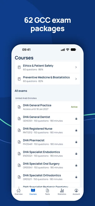
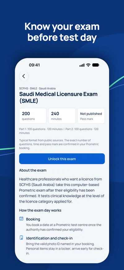
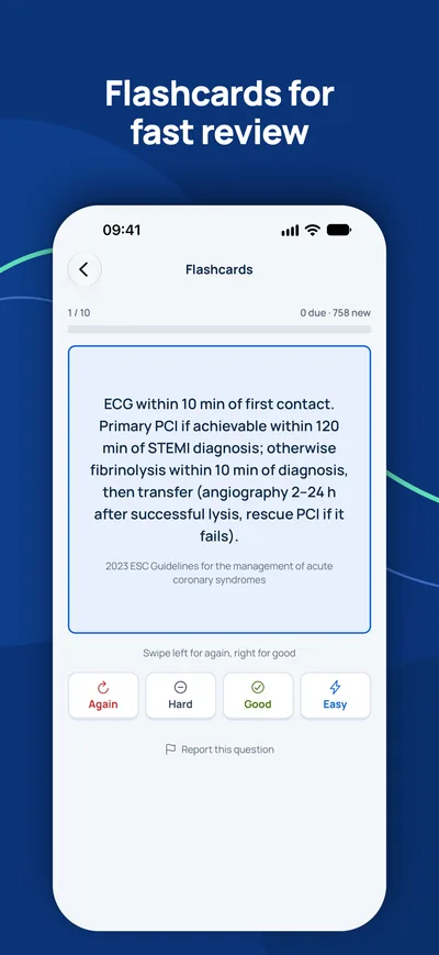
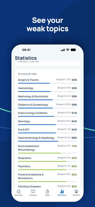
Is het examen uw enige open stap?
Controleer eerst of u het examen echt nodig hebt en welke titel u kunt verwachten. Een adviseur beantwoordt uw vragen gratis.
Geschiktheidscheck- 1Gratis adviesgesprek via video, telefoon of WhatsApp
- 2Geschiktheidscheck volgens de officiële eisen
- 3Uw plan met vaste prijzen en duidelijke stappen