
🇰🇼 MOH Kuwait
Specialist Periodontics Kuwait
Specialist dentists preparing for the Periodontics exams of DHA, DOH, Qatar DHP and SCFHS
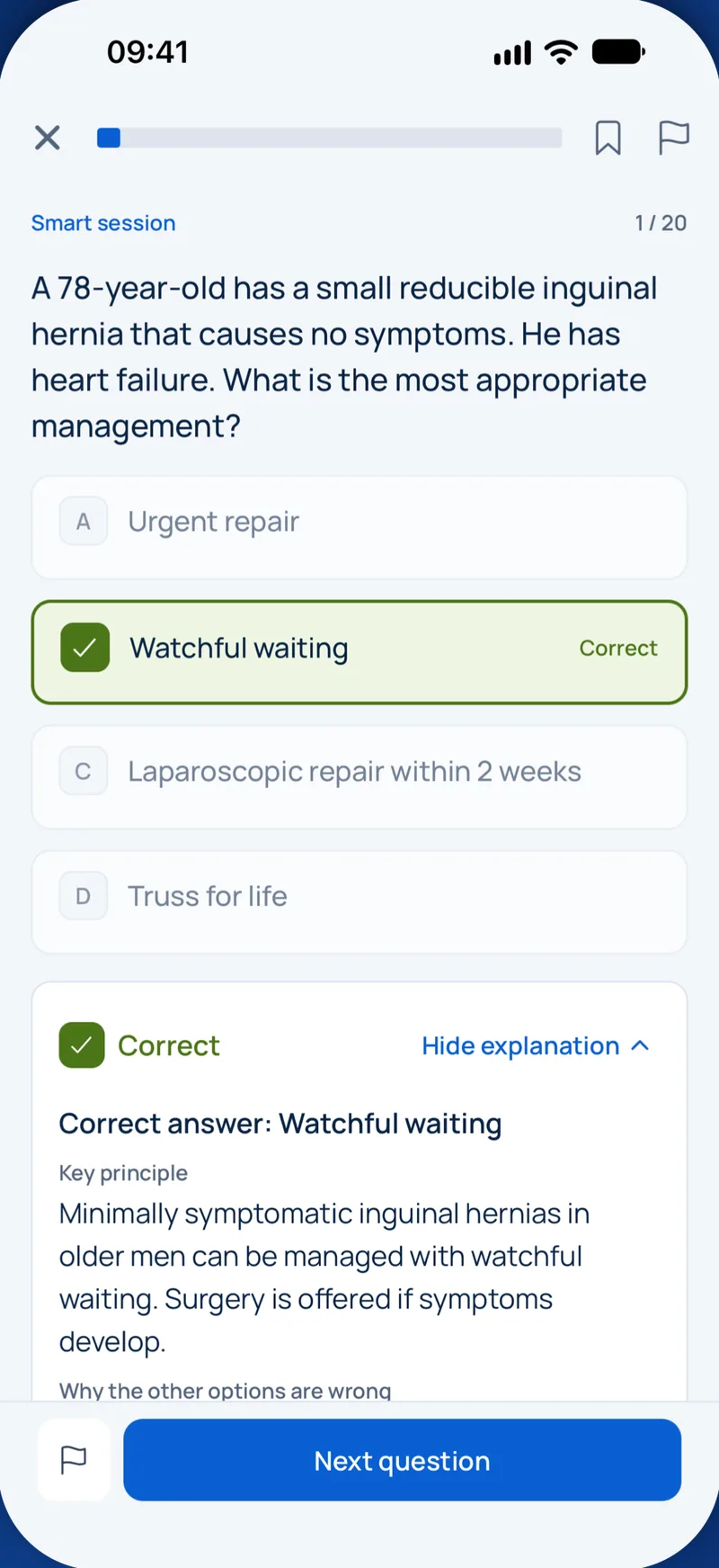

Пример вопроса
Which drug class is NOT typically associated with drug-influenced gingival enlargement?
Keys: 1–5 or A–E to choose, Enter to check and continue, F to mark.
Original MediFlow content, not official exam questions. Medical review in progress.
Syllabus and high-yield notes
1. Classification & diagnosis · 17 % вес на экзамене · 5 lessons+
- Staging (I–IV) describes severity and complexity: interdental CAL at the worst site, radiographic bone loss, tooth loss due to periodontitis, and complexity factors (PD ≥6 mm, vertical defects ≥3 mm, furcation II/III, masticatory dysfunction, fewer than 20 teeth). Complexity factors can only raise the stage.
- Grade (A–C) is assigned primarily from direct evidence of progression or, if absent, from %bone loss/age at the worst tooth (<0.25 A, 0.25–1.0 B, >1.0 C); grade B is the default, then modified by smoking (≥10 cig/day = C) and HbA1c (≥7.0% = C).
- Extent: localized if fewer than 30% of teeth are involved, generalized if 30% or more; a molar/incisor pattern is described separately and replaces the former localized aggressive periodontitis.
- A "periodontitis case" requires interdental CAL detectable at ≥2 non-adjacent teeth, or buccal/oral CAL ≥3 mm with PD >3 mm at ≥2 teeth, not attributable to non-periodontal causes.
- Gingivitis on an intact periodontium in epidemiology is BOP ≥10% with PD ≤3 mm; localized 10–30%, generalized >30%. A successfully treated periodontitis patient with BOP <10% and no PD ≥4 mm with BOP is "periodontal health on a reduced periodontium" but remains a periodontitis patient.
- Necrotizing periodontal diseases are classified by host status: chronically severely compromised (e.g., HIV/AIDS, severe malnutrition) versus temporarily or moderately compromised (e.g., stress, smoking, poor sleep).
- Gingival recession is classified by the interdental CAL (Cairo RT1–RT3), which predicts achievable root coverage: RT1 no interproximal loss, RT2 interproximal CAL ≤ buccal, RT3 interproximal CAL > buccal.
- Endo-periodontal lesions are classified 2017 by presence or absence of root damage (fracture, perforation, external resorption) and, without root damage, by periodontitis status and the extent of the deep pocket (narrow, wide, one or more surfaces).
- Periodontal health, gingivitis and the logic of the 2017 classification – In 2017 the American Academy of Periodontology (AAP) and the European Federation of Periodontology (EFP) held a World Workshop that replaced the 1999 Armitage classification. The 2018 consensus papers are now the language of every Gulf licensing exam. The biggest changes were three. First, periodontal health was defined for the first time in measurable terms. Second, the old split into chronic and aggressive periodontitis was dropped, because the two could not be separated reliably by biology. Third, every periodontitis case is now described by a stage (how severe and how complex) and a grade (how fast it progresses and how much risk the patient carries).
- Staging and grading of periodontitis step by step – Periodontitis is chronic inflammation, driven by a dysbiotic biofilm, that destroys the attachment apparatus. A patient is a periodontitis case when interdental CAL is detectable at two or more non-adjacent teeth, or when buccal or oral CAL of 3 mm or more with pocketing of more than 3 mm is present at two or more teeth, and the loss cannot be explained by other causes such as traumatic recession, caries at the cervical area, a vertical root fracture, an endodontic lesion draining through the periodontium or a third molar extraction.
- Necrotizing disease, systemic conditions, abscesses, endo-perio lesions and mucogingival conditions – Necrotizing periodontal diseases are a separate category of periodontitis because their course is acute and their link to impaired host defence is strong. Necrotizing gingivitis (NG) affects only the gingiva; necrotizing periodontitis (NP) also destroys attachment and bone; necrotizing stomatitis spreads beyond the gingiva into the mucosa and bone. The triad for diagnosis is necrosis and ulceration of interdental papillae (punched-out, often covered by a grey pseudomembrane), gingival bleeding and pain. Fetid breath, fever and lymphadenopathy are supportive. Predisposing factors include psychological stress, sleep deprivation, smoking, malnutrition and immunosuppression, especially HIV infection with low CD4 counts. The classification separates patients who are chronically severely compromised from those who are temporarily or moderately compromised, such as a student during exams.
- Periodontal structures: gingiva, periodontal ligament, cementum and alveolar bone – The periodontium is the group of tissues that attach the tooth to the jaw and protect that attachment. It has four parts: the gingiva, the periodontal ligament (PDL), the root cementum and the alveolar bone. The gingiva is the only part you see clinically; the other three are judged with the probe and the radiograph. Every diagnosis in periodontics is a statement about one or more of these tissues, so knowing their normal structure is the base for everything else in this track.
- Periodontal examination, charting and radiographic assessment – A periodontal diagnosis is only as good as the examination behind it. The classification in this topic needs specific numbers: interdental clinical attachment loss, radiographic bone loss in relation to root length, probing depths, furcation involvement, mobility, tooth loss due to periodontitis and bleeding on probing. This chapter explains how to collect those numbers correctly and how screening tools fit in.
2. Aetiology & risk · 17 % вес на экзамене · 5 lessons+
- Current pathogenesis model: polymicrobial synergy and dysbiosis (PSD). Keystone pathogens such as Porphyromonas gingivalis subvert host immunity at low abundance, and the dysregulated inflammation itself fuels the dysbiotic community.
- Bone loss is driven by the RANKL/OPG balance: Th17 cells, osteoblasts and fibroblasts express RANKL; IL-17, IL-1β, TNF-α and PGE2 increase RANKL relative to osteoprotegerin.
- Page and Schroeder lesions: initial (2–4 days, neutrophils), early (4–7 days, T lymphocytes), established (2–3 weeks, plasma cells/B cells), advanced (bone loss, transition to periodontitis).
- Smoking increases periodontitis risk dose-dependently (about 2–7-fold), masks gingival bleeding through vasoconstriction, and reduces healing after both non-surgical and surgical therapy.
- The diabetes–periodontitis link is bidirectional: poor glycaemic control raises periodontitis risk, and periodontal therapy lowers HbA1c by roughly 0.3–0.4 percentage points at 3 months.
- Advanced glycation end-products (AGEs) binding RAGE on macrophages and endothelium amplify inflammatory cytokine release and impair collagen turnover in diabetic patients.
- Local plaque-retentive factors include overhanging restorations, subgingival margins, enamel pearls, cervical enamel projections and palatogingival grooves; correcting them is part of step 1 or 2 of therapy.
- Occlusal trauma does not initiate periodontitis, but in the presence of plaque-induced inflammation jiggling forces can accelerate attachment loss (Glickman co-destruction concept vs. Waerhaug).
- The biofilm and the microbiology of periodontitis – Dental plaque is a biofilm: a structured community of microorganisms attached to the tooth and embedded in a self-produced matrix. Formation starts within minutes. Salivary proteins adsorb onto the clean enamel and form the acquired pellicle. Early colonizers, mostly Gram-positive cocci such as Streptococcus sanguinis and Actinomyces species, attach to pellicle receptors. Fusobacterium nucleatum acts as a bridge organism because it can coaggregate with both early and late colonizers. Later, Gram-negative anaerobes settle in the deeper, oxygen-poor parts of the biofilm and the pocket.
- Host response, inflammation and bone destruction – Most of the tissue damage in periodontitis is done by the host, not directly by bacteria. The bacteria start the process; an overactive or poorly controlled immune response destroys collagen and bone. Understanding the sequence of the host response helps you answer questions about cell types, cytokines and why some people break down faster than others.
- Risk factors, local factors and links to systemic disease – A risk factor is something that, if present, raises the probability of disease and, when removed, lowers it; it must be shown in longitudinal studies. A risk indicator is a plausible association from cross-sectional data. A risk marker or predictor, such as previous bone loss, signals higher risk without necessarily causing it. The two modifiable risk factors with the strongest evidence for periodontitis are smoking and diabetes, and both appear in the 2017 grading system.
- Periodontal medicine: diabetes, cardiovascular disease, pregnancy and the medically compromised patient – Periodontal medicine studies the two-way link between periodontal and systemic health. Periodontitis is a chronic inflammatory disease with an ulcerated pocket epithelium that, in generalised disease, may add up to a wound surface of several square centimetres. Bacteria and their products enter the bloodstream daily, and the inflamed tissues release cytokines such as interleukin-1, interleukin-6 and TNF-alpha, raising systemic markers like C-reactive protein. In the other direction, systemic conditions change the host response and so the course of periodontitis. For the exam you need to know where the evidence is strong (diabetes), where it is an association (cardiovascular disease, pregnancy outcomes) and how to treat medically compromised patients safely.
- Occlusal trauma, tooth mobility and local anatomical factors – Plaque causes periodontitis, but other local factors decide where and how fast it progresses. This chapter covers occlusal forces, tooth mobility, and anatomical and iatrogenic factors that make plaque control difficult or redirect the inflammatory process. These themes appear often in questions about why one tooth is worse than its neighbours.
3. Non-surgical therapy · 17 % вес на экзамене · 5 lessons+
- The EFP S3 guideline uses four steps: (1) behaviour change, supragingival plaque control and risk-factor control; (2) subgingival instrumentation with or without adjuncts; (3) treatment of non-responding sites (re-instrumentation or surgery); (4) supportive periodontal care.
- Hand and power-driven (sonic/ultrasonic) instruments are equally effective for subgingival instrumentation; quadrant-wise and full-mouth approaches give similar results.
- Systemic antibiotics (e.g., amoxicillin plus metronidazole) should not be used routinely; they may be considered for specific patient categories such as generalized stage III periodontitis in young adults with high progression rates.
- Reevaluation is performed after a healing period of about 6–12 weeks; the endpoint is pocket closure (PD ≤4 mm without BOP).
- Expected results after subgingival instrumentation: mean PD reduction about 1.5 mm and around 74% pocket closure in pockets ≥4 mm; deeper pockets show greater reduction but lower closure.
- Lasers, photodynamic therapy, local antibiotics without proven benefit, and host modulators such as NSAIDs or omega-3 are not recommended as routine adjuncts in the EFP guideline; locally delivered sustained-release chlorhexidine may be considered.
- Supportive periodontal care intervals are typically 3–12 months based on risk (e.g., PRA: BOP, residual pockets ≥5 mm, tooth loss, bone loss/age, systemic and genetic factors, smoking).
- Necrotizing gingivitis is managed by gentle debridement, chlorhexidine or hydrogen peroxide rinses, analgesia, and metronidazole only if systemic involvement (fever, lymphadenopathy) is present.
- The EFP stepwise approach: steps 1 and 2 – In 2020 the European Federation of Periodontology published an S3-level clinical practice guideline for the treatment of stage I to III periodontitis, followed in 2022 by a guideline for stage IV. S3 means the recommendations come from systematic reviews and a formal consensus process. The guideline organizes therapy into four steps that must be done in order. Each step has a goal, and the result decides whether you move on.
- Instruments and technique for subgingival instrumentation – Good instrumentation depends on knowing the instruments and holding them correctly. Every hand instrument has a handle, a shank and a working end. The terminal (lower) shank is the part closest to the blade; its position relative to the tooth surface tells you whether the blade is correctly angulated.
- Re-evaluation, special situations and supportive periodontal care – Healing after subgingival instrumentation takes time. The pocket epithelium reattaches within one to two weeks, but connective tissue maturation and shrinkage continue for months. Re-evaluation is therefore done about 6 to 12 weeks after step 2, most commonly at around 3 months. Re-evaluation includes a full periodontal chart with PD, CAL, BOP, mobility and furcations, plus plaque score and a review of risk factors.
- Periodontal pharmacotherapy: antiseptics, antibiotics, host modulation and pain control – Drugs in periodontics are adjuncts. The cause of periodontitis is a biofilm that is protected against antimicrobials by its matrix, so mechanical disruption by the patient and the clinician always comes first. Antimicrobials can help in selected situations, but each use must be weighed against side effects and antimicrobial resistance. The EFP S3 guideline (2020) gives clear recommendations that examiners like to test.
- Patient safety, ethics and professionalism in periodontics – The Qatar specialty blueprint gives patient safety and professionalism and ethics 5 percent each, the same weight as whole clinical areas. Many questions are scenarios: what should you do first, who should you tell, what must you record. The answers follow a few principles that the WHO Patient Safety Curriculum and regional codes such as the SCFHS Professionalism and Ethics handbook share: respect autonomy, act in the patient's best interest, avoid harm, be honest, be fair, and work within your competence.
4. Surgical therapy · 17 % вес на экзамене · 5 lessons+
- Step 3 surgery targets residual pockets ≥6 mm after step 2; access flap surgery yields greater PD reduction than re-instrumentation in deep pockets, at the cost of more recession.
- Critical probing depth (Lindhe 1982): for non-surgical therapy about 2.9 mm and for modified Widman flap about 4.2 mm; instrumenting shallower sites causes attachment loss.
- Modified Widman flap: internal bevel incision 0.5–1 mm from the margin, sulcular incision, horizontal incision, minimal bone recontouring, flap repositioned for close adaptation; aims at healing with long junctional epithelium.
- Apically positioned flap eliminates pockets while preserving keratinized tissue; it requires a split or full-thickness flap with vertical releasing incisions and is not used on the palate.
- Osseous resective surgery: osteoplasty reshapes bone without removing supporting bone; ostectomy removes supporting bone to establish positive architecture.
- Furcation degree II/III in maxillary molars: periodontal surgery (regeneration where possible for degree II) is recommended; root resection, tunnelling and extraction are options. Mandibular degree II is the best indication for regeneration.
- Crown lengthening must re-establish a supracrestal tissue attachment (former biologic width) of about 2 mm plus sulcus, i.e., typically ≥3 mm from the planned restoration margin to the bone crest.
- Distal wedge procedures remove fibrous tissue distal to terminal molars; maxillary tuberosity usually triangular/square incisions, mandibular retromolar linear incisions following the ridge.
- Indications for periodontal surgery and access flaps – Periodontal surgery belongs to step 3 of therapy. It is never a substitute for steps 1 and 2. The classic indication is a residual pocket of 6 mm or more with bleeding at re-evaluation, in a patient with good plaque control (plaque score around 20% or less) and controlled risk factors. Surgery gives the clinician direct vision of the root and bone, which matters because the effectiveness of closed instrumentation falls as pockets get deeper and in furcations.
- Resective surgery: osseous surgery, furcations and crown lengthening – Resective periodontal surgery aims to eliminate pockets by bringing the soft tissue and bone into a shape that the patient can clean. The ideal is positive architecture: the interdental bone is more coronal than the radicular (buccal and lingual) bone, following the scalloped contour of the CEJ. Periodontitis often creates negative or flat architecture, craters and ledges, which keep soft tissue pockets after healing.
- Suturing, healing and post-operative care – Suturing holds the flap in its planned position until early healing has stabilized it. The choice of suture technique depends on whether buccal and lingual flaps are moved together or separately and on the desired position of the flap.
- Complications of periodontal surgery: prevention and management – Surgical techniques and complications carry 14 percent of the periodontics blueprint. Complications are best managed by preventing them: a full medical history, knowledge of anatomy, a flap design that keeps blood supply, careful handling of soft tissue and clear post-operative instructions. When something does go wrong, the examiner wants a calm, ordered response.
- Periodontics and other specialties: restorative, orthodontic and endodontic interrelations – Periodontal health is the foundation for almost all other dental treatment, and treatment in other specialties can damage the periodontium. The Qatar blueprint gives this interrelation 7 percent. The questions usually ask about sequence (which treatment first), restorative margins, orthodontics in periodontally compromised patients and endodontic-periodontal lesions.
5. Regeneration & plastic surgery · 16 % вес на экзамене · 5 lessons+
- True periodontal regeneration means new cementum, functionally oriented PDL and alveolar bone on a previously diseased root surface; it can only be proven histologically.
- Intrabony defects ≥3 mm deep are indications for regeneration (EMD or GTR, with or without graft); narrow defect angles (<25°) and more walls predict better outcomes.
- Papilla preservation flaps: modified (Cortellini) for interdental spaces >2 mm, simplified (SPPF) for ≤2 mm; minimally invasive surgical technique (MIST/M-MIST) limits flap elevation.
- Melcher’s concept: the cell type that repopulates the root surface determines healing; GTR excludes epithelium and gingival connective tissue so PDL cells can repopulate.
- Enamel matrix derivative (amelogenins) promotes cementum formation and mimics root development; EFP recommends EMD or GTR for deep intrabony defects.
- Coronally advanced flap plus connective tissue graft is the gold standard for root coverage of single and multiple RT1 recessions; RT2 allows partial coverage; RT3 rarely full coverage.
- Free gingival grafts increase keratinized tissue width reliably but have poor colour match; they are mainly used apical to recessions or around implants/prosthetic sites.
- Smoking reduces regeneration and root coverage outcomes; patient factors (plaque control, smoking), defect factors and surgical factors must all be optimized.
- Principles of periodontal regeneration and case selection – Repair and regeneration are different outcomes. Repair is healing that restores continuity but not the original architecture, typically a long junctional epithelium along the root or scar tissue. Regeneration is the reproduction of the lost tissues: new cementum, a functionally oriented periodontal ligament and new alveolar bone. New attachment means new connective tissue fibres inserted into new cementum on a root surface that had lost attachment. True regeneration can only be proven histologically; clinically we measure CAL gain and radiographic bone fill.
- Materials, flap designs and furcation regeneration – Regenerative materials fall into barrier membranes, biologic agents and bone substitutes. Three terms describe what a graft can do. Osteogenesis means the graft contains living cells that form bone. Osteoinduction means the graft contains signals, such as bone morphogenetic proteins (BMPs), that recruit and differentiate stem cells into bone-forming cells. Osteoconduction means the graft serves as a scaffold along which bone grows.
- Periodontal plastic surgery: recession, phenotype and frenum – Periodontal plastic surgery corrects defects of gingiva, alveolar mucosa and bone that are caused by anatomy, development, trauma or disease. The most common problem is gingival recession, the apical shift of the gingival margin beyond the CEJ. Causes include a thin phenotype, traumatic brushing, labial tooth position, orthodontic movement outside the bony housing, frenum pull and periodontitis.
- Site preparation: ridge preservation, bone augmentation and sinus floor elevation – Pre-surgical assessment and site preparation for implants carry 14 percent of the periodontics blueprint. The principles of periodontal regeneration from the earlier chapters (space maintenance, blood clot stability, cell exclusion, primary closure) also govern bone augmentation. The difference is that there is no tooth and no periodontal ligament: we are building bone for an implant, not new attachment on a root.
- Peri-implant soft and hard tissue management – Peri-implant soft and hard tissue management carries 8 percent of the blueprint. The soft tissue around an implant is different from gingiva: collagen fibres run parallel to the implant surface rather than inserting into it, there is no PDL, and blood supply comes only from the periosteum and the supracrestal vessels. This makes the soft tissue seal weaker and less vascular, and explains why tissue thickness and keratinised mucosa matter for long-term stability and aesthetics.
6. Implants & peri-implant disease · 16 % вес на экзамене · 5 lessons+
- Peri-implant mucositis: bleeding and/or suppuration on gentle probing without bone loss beyond initial remodelling; it is reversible.
- Peri-implantitis case definition: BOP/suppuration, increased probing depth compared with previous examinations, and progressive bone loss beyond initial remodelling. Without baseline data: BOP plus PD ≥6 mm and bone level ≥3 mm apical to the most coronal intraosseous part.
- History of periodontitis, poor plaque control and lack of regular maintenance are strong risk indicators for peri-implantitis; smoking and diabetes have less conclusive evidence.
- Peri-implant tissues lack a PDL and cementum; connective tissue fibres run parallel to the implant surface and blood supply comes from periosteal and bone vessels, making peri-implant lesions larger and progress faster than periodontitis lesions.
- Treatment of peri-implantitis follows a stepwise approach: non-surgical therapy first, then surgical access (resective, reconstructive in contained defects), with prosthesis modification for cleansability.
- Implant placement timing: type 1 immediate, type 2 early with soft-tissue healing (4–8 weeks), type 3 early with partial bone healing (12–16 weeks), type 4 late (≥6 months).
- Minimum distances: about 1.5 mm between implant and tooth, 3 mm between adjacent implants, and adequate buccal bone (≥2 mm ideally) to reduce recession.
- Excess cement, poor prosthetic design and lack of keratinized mucosa are local factors linked to peri-implant inflammation.
- Peri-implant tissues and implant planning – An osseointegrated implant is anchored by direct bone-to-implant contact, without a periodontal ligament. This single difference explains most clinical differences between teeth and implants: implants cannot move physiologically, have no ligament proprioception, and have no ligament blood supply or cells that can regenerate attachment.
- Peri-implant health, mucositis and peri-implantitis: diagnosis – The 2017 World Workshop defined peri-implant conditions in parallel with periodontal ones. The central idea is comparison with a baseline. After placement and loading, crestal bone remodels; this initial remodelling is physiological. A radiograph and probing depth recorded after the prosthesis is in place (at the end of remodelling, usually at loading or about one year later) are the baseline against which every later change is judged.
- Prevention and treatment of peri-implant diseases – In 2023 the EFP published an S3-level guideline for the prevention and treatment of peri-implant diseases. It follows the same stepwise logic as periodontitis therapy: control risk factors and biofilm first, then non-surgical treatment, then surgery for persisting disease, followed by lifelong supportive care.
- Implant indications, contraindications, osseointegration and biomechanics – This section of the blueprint was the least covered in our bank, yet it is the logical first step of implant care: deciding whether a patient should receive an implant at all, and understanding what holds an implant in bone and what forces it must resist.
- Pre-surgical planning, timing of placement and loading, and implant maintenance – Good implant outcomes are decided before the first incision. Planning starts from the restoration that the patient needs and works backwards to the implant position and the bone and soft tissue required (prosthetically driven or restoration-driven planning). After placement, lifelong supportive care decides whether the implant stays healthy.
Classification & diagnosis
Lesson 1Periodontal health, gingivitis and the logic of the 2017 classification
In 2017 the American Academy of Periodontology (AAP) and the European Federation of Periodontology (EFP) held a World Workshop that replaced the 1999 Armitage classification. The 2018 consensus papers are now the language of every Gulf licensing exam. The biggest changes were three. First, periodontal health was defined for the first time in measurable terms. Second, the old split into chronic and aggressive periodontitis was dropped, because the two could not be separated reliably by biology. Third, every periodontitis case is now described by a stage (how severe and how complex) and a grade (how fast it progresses and how much risk the patient carries).
The classification has four big chapters: periodontal health, gingival diseases and conditions; periodontitis (including necrotizing periodontal diseases and periodontitis as a manifestation of systemic disease); other conditions affecting the periodontium (systemic diseases, abscesses, endo-periodontal lesions, mucogingival conditions, traumatic occlusal forces, tooth- and prosthesis-related factors); and peri-implant diseases and conditions.
Three terms are needed before anything else. Probing depth (PD) is the distance from the gingival margin to the base of the pocket. Clinical attachment level (CAL) is the distance from the cementoenamel junction (CEJ) to the base of the pocket; it measures how much attachment has been lost, independent of where the gingival margin sits. Bleeding on probing (BOP) is bleeding within about 30 seconds of gentle probing (around 0.25 N) and is the key clinical sign of inflammation. A reduced periodontium means there has been attachment loss in the past, either from periodontitis or from non-periodontitis causes such as recession or crown lengthening.
| Situation | Clinical definition (case level) |
|---|---|
| Clinical health, intact periodontium | PD 3 mm or less, BOP under 10% of sites, no attachment or bone loss |
| Clinical health, reduced periodontium, non-periodontitis patient | PD 3 mm or less, BOP under 10%, attachment loss present (for example from recession) |
| Stable periodontitis patient (reduced periodontium) | PD 4 mm or less, no site of 4 mm with BOP, BOP under 10% |
| Gingivitis, intact or reduced periodontium | BOP 10% or more, PD 3 mm or less (or no new attachment loss in treated periodontitis) |
| Localized gingivitis | BOP 10 to 30% of sites |
| Generalized gingivitis | BOP more than 30% of sites |
A crucial teaching point: a patient who has once had periodontitis remains a periodontitis patient for life. Even when treatment succeeds, the diagnosis is not changed to health. Instead the patient is described as a stable periodontitis patient, and the stage at diagnosis stays on record. If such a patient has BOP of 10% or more but no deep pockets, the label is gingival inflammation in a stable periodontitis patient. A site of 4 mm with bleeding, or any site of 5 mm or more, is a pocket that is not closed and counts as unstable.
Gingival diseases are divided into dental biofilm-induced gingivitis and non-biofilm-induced gingival conditions. Biofilm-induced gingivitis can be modified by systemic factors (sex steroid hormones in puberty, menstruation, pregnancy and oral contraceptives; hyperglycaemia; leukaemia; smoking; malnutrition) and by local factors (restoration margins, hyposalivation). Drug-influenced gingival enlargement is part of this group. The three drug classes to remember are anticonvulsants (phenytoin), the immunosuppressant ciclosporin and calcium channel blockers (nifedipine, amlodipine, verapamil, diltiazem). Plaque control reduces the enlargement, and substitution of the drug by the physician is the definitive step when possible.
Pregnancy gingivitis is common in the second and third trimester because oestrogen and progesterone increase vascular permeability and change the host response. A localized, pedunculated, easily bleeding red mass on an interdental papilla in pregnancy is a pyogenic granuloma, often called pregnancy tumour or epulis gravidarum. It is managed with meticulous plaque control and usually observed until after delivery, because many shrink; excision is reserved for lesions that bleed, interfere with eating or persist post partum.
Non-biofilm-induced conditions include genetic disorders (hereditary gingival fibromatosis), specific infections (herpetic gingivostomatitis, candidiasis, gonococcal and syphilitic lesions), inflammatory and immune conditions (lichen planus, pemphigus vulgaris, mucous membrane pemphigoid, lupus), reactive lesions, neoplasms, endocrine and nutritional causes (scurvy) and traumatic lesions. Desquamative gingivitis is a clinical description, not a diagnosis; biopsy with direct immunofluorescence identifies the underlying disease.
Sources: Chapple ILC, Mealey BL et al. Periodontal health and gingival diseases and conditions on an intact and a reduced periodontium. J Periodontol 2018;89(Suppl 1):S74-S84 · Caton JG, Armitage G, Berglundh T et al. A new classification scheme for periodontal and peri-implant diseases and conditions. J Clin Periodontol 2018;45(Suppl 20):S1-S8 · Newman and Carranza's Clinical Periodontology and Implantology, 14th ed. Elsevier 2023
Об экзамене MOH Kuwait Specialist Periodontics
Сколько вопросов на экзамене MOH Kuwait Specialist Periodontics?+
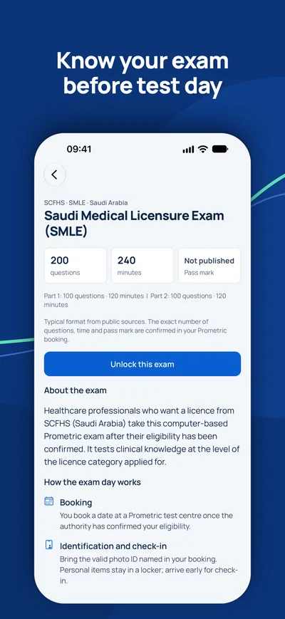
не опубликовано. Точное число вопросов, время и проходной балл подтверждаются при записи.
Какие темы весят больше всего?+
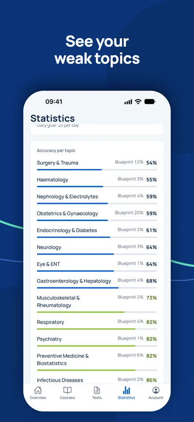
Крупнейшие темы: Classification & diagnosis (17 %), Aetiology & risk (17 %), Non-surgical therapy (17 %). Курс охватывает 6 тем и 30 глав.
Как подготовиться к экзамену MOH Kuwait Specialist Periodontics?+
Проходите главы тема за темой, повторяйте ключевые заметки, затем тренируйтесь на пробных экзаменах с таймером в формате вашего органа.
Вопросы
Как долго действует доступ?+
Доступ действует 90 дней с момента оплаты для выбранной страны и профессии.
Какие устройства можно использовать?+
Любой браузер на компьютере, планшете или телефоне, а также приложение MediFlow с тем же аккаунтом.
Можно ли вернуть деньги?+
Пожалуйста, прочитайте правила возврата перед покупкой. Правила возврата

iPhone · iPad
GCC Medical Exam Prep
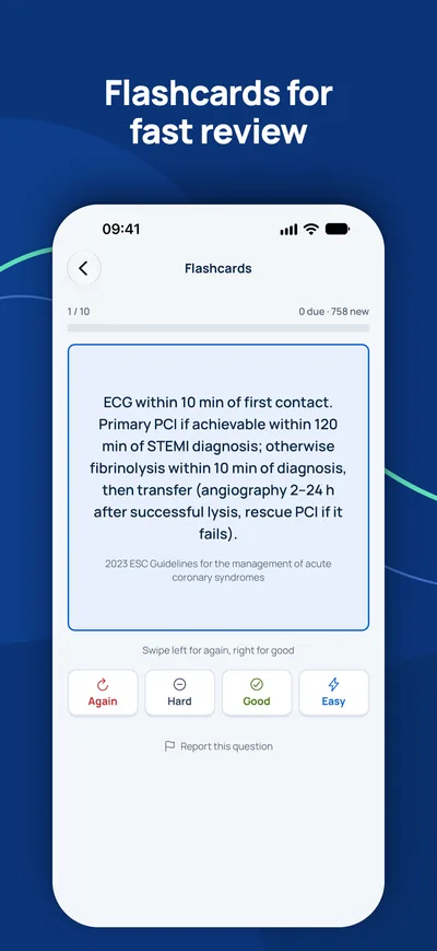
Готовьтесь на iPhone к лицензионным экзаменам DHA, DOH, MOHAP, SCFHS и другим экзаменам стран Залива.

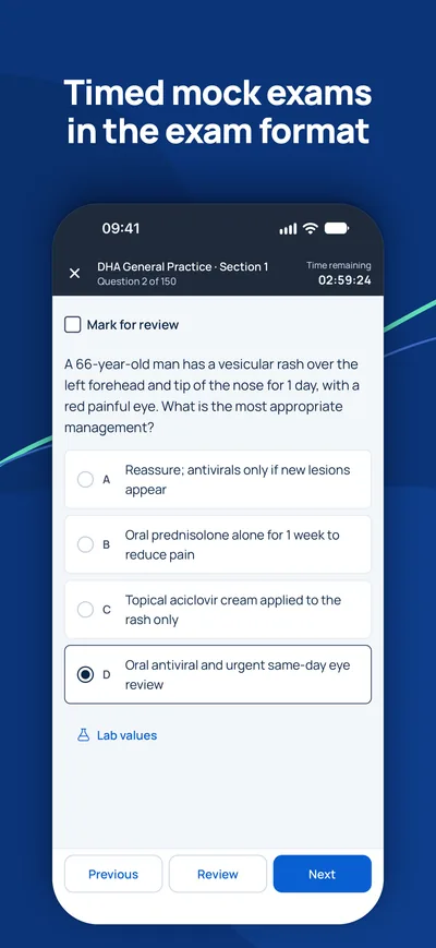
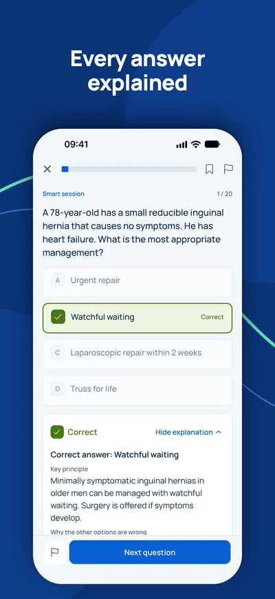
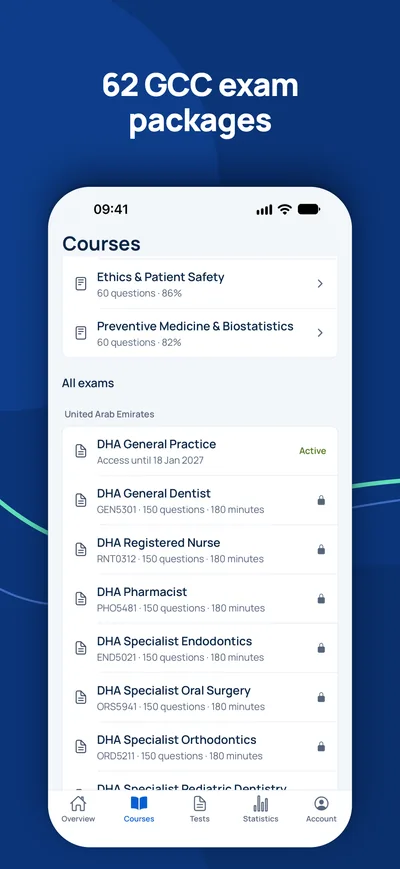
Экзамен — ваш единственный оставшийся шаг?
Сначала проверьте, нужен ли вам экзамен и на какое звание вы можете рассчитывать. Консультант ответит бесплатно.
Проверка соответствия- 1Бесплатная консультация по видео, телефону или WhatsApp
- 2Проверка соответствия официальным требованиям
- 3Ваш план с фиксированными ценами и понятными шагами