
🇶🇦 DHP
Specialist Paediatric dentistry Qatar
Specialist dentists preparing for the Paediatric dentistry exams of DHA, DOH, Qatar DHP and SCFHS
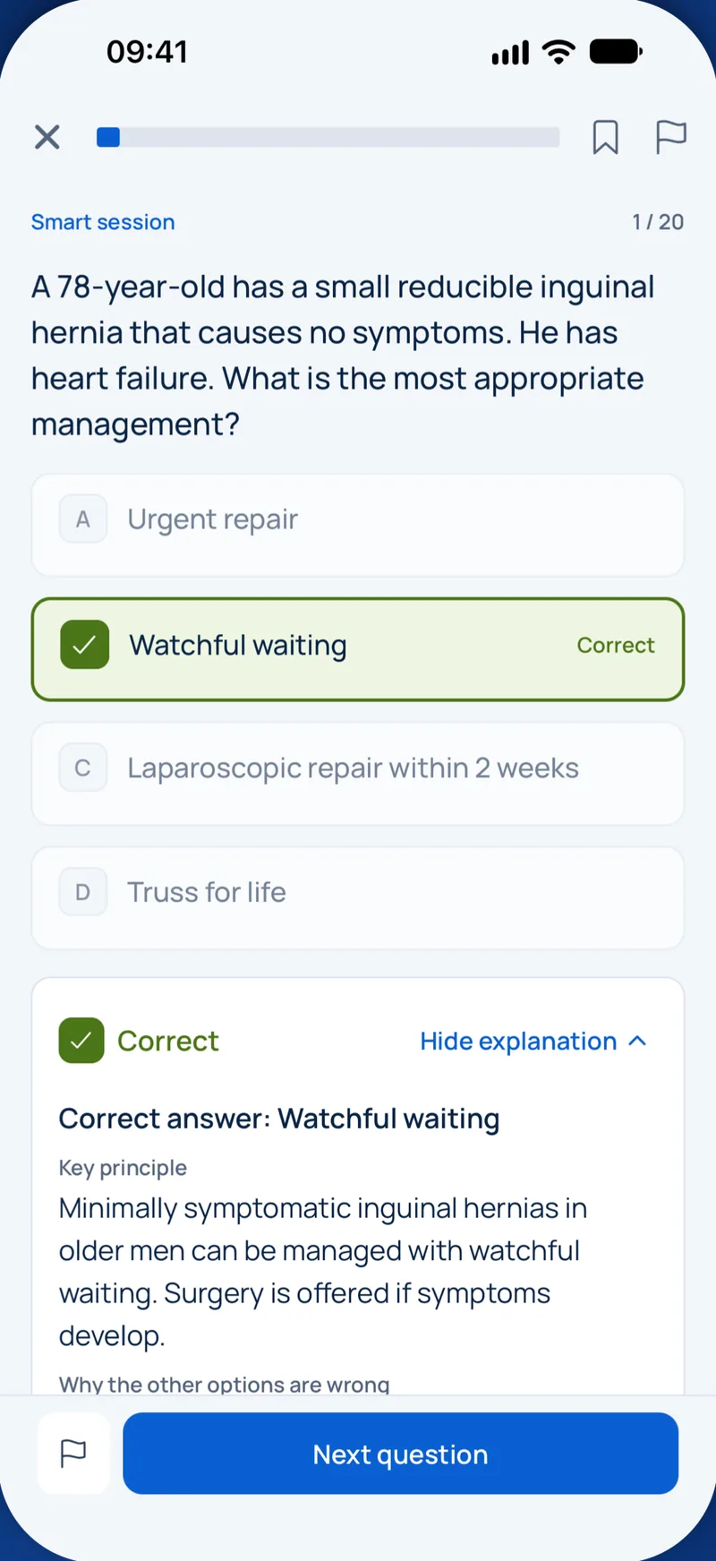
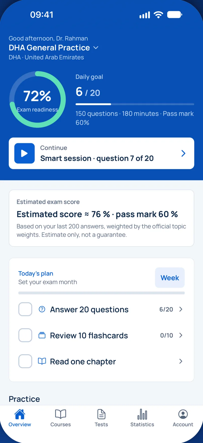
Пример вопроса
At about what stage of intrauterine life does calcification of the primary teeth begin?
Keys: 1–5 or A–E to choose, Enter to check and continue, F to mark.
Original MediFlow content, not official exam questions. Medical review in progress.
Syllabus and high-yield notes
1. Growth & development · 15 % вес на экзамене · 5 lessons+
- The leeway space (Proffit) averages about 1.5 mm per side in the maxilla and 2.5 mm per side in the mandible; preserving it with a passive lingual arch can resolve mild mandibular crowding.
- Primate spaces lie mesial to the maxillary primary canines and distal to the mandibular primary canines and allow the early mesial shift of the first permanent molars.
- A distal step of the primary second molars predicts a Class II molar relationship; a flush terminal plane may become Class I or end-on depending on space and growth.
- The 'ugly duckling' stage (median diastema and distally flared lateral incisors at 8–11 years) is physiological and usually closes as the canines erupt.
- Choose the space maintainer by the tooth lost and the stage: band-and-loop for a single primary first molar, distal shoe if the second primary molar is lost before the first permanent molar erupts, lingual holding arch for bilateral mandibular loss after incisor eruption.
- Ectopic eruption of the maxillary first permanent molar self-corrects ('jumps') in many cases; an irreversible lock is treated by distal tipping with a separator, brass wire or a Halterman appliance.
- Cervical vertebral maturation stage CS3 indicates that the pubertal growth peak is expected within about a year, the best time for functional Class II treatment.
- Syndromic clues: hypodontia with conical teeth and heat intolerance suggests hypohidrotic ectodermal dysplasia; multiple supernumeraries with delayed eruption and aplastic clavicles suggest cleidocranial dysplasia (RUNX2).
- Tooth formation, eruption timing and developmental anomalies – Every question about a child's dentition starts with one idea: teeth form on a timetable. If you know when crowns calcify, when teeth erupt and when roots close, you can date a defect, predict a problem and choose a treatment that respects the stage of development. This chapter builds that timetable and then shows how inherited and acquired disturbances change it.
- Occlusal development from the primary to the permanent dentition – Space is the currency of the developing dentition. Permanent incisors are larger than the primary incisors they replace, while permanent canines and premolars are together smaller than the primary canine and molars. Growth, spacing and the order in which teeth erupt decide whether the arch has enough room. Understanding these numbers lets you tell a normal transitional stage from a real problem.
- Space management and interceptive treatment of eruption problems – When a primary molar is lost early, the teeth around the gap drift. The first permanent molar moves mesially and tips, and the space for the premolar shrinks. Space loss is fastest in the first months after extraction, is greater in the maxilla, and is greater after loss of a primary second molar than a first molar. A space maintainer is a passive appliance that holds the gap until the successor erupts. Before placing one, confirm on a radiograph that the successor is present and not about to erupt; if more than about half of its root is formed and only a thin layer of bone covers it, eruption is near and maintenance may be unnecessary.
- Oral habits, crossbites and mixed dentition analysis – The mixed dentition period, from about 6 to 12 years, is when many malocclusions first show and when simple treatment can prevent bigger problems. The paediatric dentist does not need to be an orthodontist, but must recognise which problems need early interception, which can wait, and which need referral. This chapter covers non-nutritive sucking habits, anterior and posterior crossbites, and the space analyses used to predict crowding.
- Oral lesions of infants and children – Many oral lesions in children are harmless and self-limiting, but they worry parents and some need treatment or referral. The paediatric dentist should be able to name the common lesions of newborns, infants and older children, explain their cause, and decide whether to observe, treat or refer. This chapter groups them by age and by site.
2. Behaviour & sedation · 15 % вес на экзамене · 5 lessons+
- Oral midazolam for paediatric dental sedation is typically 0.25–0.5 mg/kg (maximum about 20 mg); its effect peaks after roughly 20–30 minutes and lasts under an hour.
- Reversal agents: flumazenil 0.01 mg/kg IV (max 0.2 mg per dose) for benzodiazepines; naloxone 0.1 mg/kg (max 2 mg) for full opioid reversal. Monitor afterwards because re-sedation can occur.
- Deep sedation requires an independent observer whose only task is monitoring and who is skilled in paediatric airway management, plus capnography.
- Nitrous oxide is contraindicated or used with caution in vitamin B12 deficiency, MTHFR deficiency, recent middle-ear surgery, pneumothorax, bleomycin therapy and the first trimester; routine concentration should not exceed 50 %.
- Discharge after sedation needs return to near baseline: patent airway, stable vital signs, can talk or sit as appropriate for age, adequate hydration, and the child stays awake at least 20 minutes in a quiet environment.
- Protective stabilisation needs documented informed consent, a clear indication and the least restrictive method; it is never used as punishment or for staff convenience.
- Laryngospasm: apply jaw thrust and positive-pressure 100 % oxygen first; if it persists, a small dose of succinylcholine with intubation readiness is required.
- Office sedation is generally limited to ASA I–II children; ASA III and above, or children with difficult airways, are better treated in a hospital setting.
- Child development, anxiety assessment and non-pharmacological behaviour guidance – Behaviour guidance is the continuous interaction between the dental team, the child and the parent that aims to build trust, reduce fear and deliver safe, good quality care. It is not a set of tricks for forcing a child to sit still. The American Academy of Pediatric Dentistry (AAPD) describes it as a continuum: most children respond to communication alone, a smaller group needs pharmacological help, and a few need general anaesthesia. Choosing well depends on understanding how children think at each age and on measuring how anxious or cooperative a child is.
- Sedation in children: patient selection, nitrous oxide and oral midazolam – Sedation reduces anxiety and movement so that treatment can be completed safely, but it is a continuum. A drug given for minimal sedation can produce deep sedation or even general anaesthesia in a susceptible child. The AAP/AAPD guideline (Coté and Wilson, 2019) therefore requires that the team be able to rescue a patient from one level deeper than the level intended.
- Monitoring, sedation emergencies, reversal and discharge – Most serious sedation incidents in children start as an airway problem that was noticed late. The answer is a monitoring standard matched to the intended depth, trained people with clear roles, emergency equipment sized for children, and a practised plan. The AAP/AAPD guideline uses the acronym SOAPME for preparation: Suction, Oxygen, Airway equipment of the right size, Pharmacy (resuscitation and reversal drugs), Monitors, and special Equipment such as a defibrillator.
- Local anaesthesia in children: doses, techniques and complications – Good local anaesthesia is the foundation of behaviour guidance. A child who feels pain during treatment learns that the dental chair hurts, and that lesson is hard to undo. Most problems with local anaesthesia in children come from three sources: giving too much drug for the child's weight, using adult anatomy for the injection site, and forgetting that soft tissue numbness lasts longer than the procedure.
- General anaesthesia for dental treatment in children – Some children cannot be treated safely in the dental chair, even with the best behaviour guidance and sedation. For them, general anaesthesia (GA) allows all treatment to be completed in one session under controlled conditions. GA is never a first choice for convenience. It carries small but real risks, needs a hospital or accredited facility, and does not by itself change the habits that caused the disease. This chapter explains when GA is justified, how the child is prepared, how the treatment plan differs, and what happens afterwards.
3. Caries & prevention · 20 % вес на экзамене · 5 lessons+
- Severe early childhood caries (S-ECC) is any smooth-surface caries in a child younger than 3 years; from 3 to 5 years it includes cavitated, missing or filled smooth surfaces in maxillary anterior teeth or dmfs thresholds of ≥4 (age 3), ≥5 (age 4), ≥6 (age 5).
- 38 % silver diamine fluoride contains about 44,800 ppm fluoride; twice-yearly application arrests more lesions than annual application. Warn families about permanent black staining of the lesion.
- 5 % sodium fluoride varnish contains 22,600 ppm fluoride and is applied every 3–6 months according to caries risk.
- Non-cavitated proximal lesions in enamel or the outer third of dentine can be arrested micro-invasively by resin infiltration, avoiding restorations.
- In deep lesions of vital, asymptomatic teeth, selective removal to soft dentine over the pulp with hard dentine at the periphery reduces pulp exposure without worsening outcomes.
- MIH: demarcated opacities on one to four first permanent molars, often with incisor involvement; severely affected hypersensitive molars are best protected with preformed metal crowns.
- Sugar frequency matters more than quantity; the WHO advises free sugars below 10 % of energy intake, ideally below 5 %.
- Fluoride works mainly topically after eruption: it inhibits demineralisation, enhances remineralisation and inhibits bacterial enolase.
- How caries develops in children and how to assess risk – Dental caries is a dynamic, biofilm-mediated, sugar-driven disease that causes a net loss of mineral from the hard tissues. Bacteria in plaque ferment dietary sugars to acids; when the pH at the tooth surface falls, enamel loses mineral (demineralisation); when the pH recovers, saliva and fluoride allow mineral to return (remineralisation). A lesion forms only when, over time, loss outweighs gain. This balance view explains why prevention works: every factor that shortens acid attacks or speeds repair tips the balance back.
- Fluoride, silver diamine fluoride and other preventive measures – Fluoride is the cornerstone of caries prevention. For a long time it was thought to work mainly by being built into developing enamel before eruption. We now know that its main effect is topical and after eruption. Low, constant concentrations of fluoride in saliva and plaque fluid are what protect the tooth.
- Detecting lesions and minimally invasive management, including MIH – Modern caries management asks two questions about each lesion: is it active or arrested, and is it cavitated or not? Non-cavitated lesions can usually be arrested without a drill. Cavitated lesions that cannot be cleaned by the patient usually need a restoration or sealing. Accurate detection is therefore the first step.
- Restoring primary teeth: materials, crowns and the Hall technique – Restoring a primary tooth is different from restoring a permanent one. The tooth only needs to last until it exfoliates, the enamel and dentine are thinner, the pulp is large and close to the surface, and the child may tolerate only a short procedure. The best restoration is therefore the one that survives until natural exfoliation with the least burden for the child. This chapter compares the main options and explains how to choose between them.
- Anticipatory guidance, diet counselling and the dental home – Anticipatory guidance means telling parents about expected changes before they happen, so that they can prevent problems instead of reacting to them. In paediatric dentistry, this covers feeding, toothbrushing, fluoride, teething, oral habits, injury prevention and diet at each age. It is most effective when it starts early and is repeated at every visit. The idea behind the dental home is that every child should have one regular source of dental care, ideally established by 12 months of age, where prevention and treatment are coordinated.
4. Pulp therapy · 20 % вес на экзамене · 5 lessons+
- For primary molars with deep caries and no signs of irreversible pulpitis, indirect pulp treatment has higher long-term success than pulpotomy (AAPD 2017 guideline).
- Recommended pulpotomy medicaments in primary molars are MTA (preferred) and formocresol; ferric sulfate 15.5 % and calcium hydroxide show lower success, the latter with internal resorption.
- Primary root canals are filled with resorbable materials: ZOE or iodoform–calcium hydroxide pastes. A stainless steel crown afterwards improves the outcome.
- Extraction rather than pulp therapy is indicated when a primary tooth has pathological resorption of more than about one-third of the root, internal resorption, or a non-restorable crown.
- In immature permanent teeth, vital pulp therapy (partial or full pulpotomy with a calcium silicate cement) allows continued root development, even when symptoms suggest irreversible pulpitis (ESE 2019, AAE 2021).
- Regenerative endodontics (AAE): first visit 1.5 % NaOCl (20 mL per canal) and a low-concentration antibiotic paste or calcium hydroxide; second visit 17 % EDTA, induced bleeding to the CEJ, collagen matrix and calcium silicate barrier.
- Grey MTA and bismuth-oxide-containing materials discolour teeth; in aesthetic zones use bismuth-free calcium silicate cements.
- Electric and thermal pulp tests are unreliable in primary teeth and young permanent teeth; base the diagnosis on pain history, clinical signs and radiographs.
- Pulp biology, diagnosis and case selection in children – Pulp therapy in children is not adult endodontics on smaller teeth. Primary teeth have a limited lifespan, a permanent successor sitting under their roots, and a pulp that reacts differently to caries. Young permanent teeth have wide open apices, thin dentine walls and an excellent blood supply. Every decision in this topic therefore starts with three questions: what is the pulp status, how much longer must the tooth stay, and can it be restored with a good coronal seal?
- Vital and non-vital pulp therapy in primary teeth – Pulp therapy in primary teeth follows a ladder. The least invasive treatment that keeps the tooth healthy until exfoliation is the right one. In order: indirect pulp treatment, direct pulp capping (rarely), pulpotomy, pulpectomy, and extraction.
- Pulp therapy for immature permanent teeth – An immature permanent tooth has an open apex and thin, short roots. Root development continues for about three years after eruption, and it only continues if the pulp stays alive. The goal is therefore to keep the pulp vital whenever possible (apexogenesis, meaning continued physiological root formation). If the pulp is already necrotic, the choices are regenerative endodontics or apexification.
- Odontogenic infection, antibiotics and pain control in children – Pulp disease in a child often ends as an infection: a dentoalveolar abscess, a sinus tract, or a facial swelling. Most of these infections are treated by removing the source, either by pulp therapy or extraction. Antibiotics have a limited, specific role. Because children have less physiological reserve than adults, a spreading facial infection can become dangerous quickly. This chapter explains how to recognise severity, when antibiotics are and are not indicated, and how to control pain safely.
- Outcomes, follow-up and failure of pulp therapy – A pulp treatment is only successful if the tooth stays healthy until it is lost naturally or, for a permanent tooth, for life. Success is judged clinically and radiographically over time, not on the day of treatment. This chapter explains how success is defined, what the evidence shows for the main techniques, how to recognise failure, and what to do when it happens.
5. Trauma · 20 % вес на экзамене · 5 lessons+
- IADT 2020 splint times: subluxation (if needed for comfort), extrusion and avulsion 2 weeks; lateral luxation, intrusion (when repositioned), alveolar fracture and root fracture 4 weeks; cervical-third root fracture up to 4 months.
- In mature avulsed and replanted teeth, start root canal treatment within 2 weeks, before splint removal if possible; immature teeth are monitored for revascularisation.
- Intruded immature permanent teeth are allowed to re-erupt spontaneously regardless of the degree; mature teeth intruded more than 7 mm are repositioned surgically.
- Best storage media for an avulsed tooth are HBSS or milk, then saliva or saline; tap water is harmful to PDL cells.
- After avulsion, systemic antibiotics are optional; under 12 years use phenoxymethylpenicillin or amoxicillin rather than tetracyclines. Check tetanus status.
- In growing children, an ankylosed incisor in infraposition is decoronated to preserve alveolar bone width and height for later implant or prosthetic replacement.
- In primary teeth, the main goal is to protect the developing successor; a displaced coronal fragment after root fracture is removed while the apical fragment is left to resorb.
- Pulp canal obliteration after luxation is a sign of pulp survival and does not require root canal treatment unless periapical pathology develops.
- Assessment and fractures of permanent teeth – Dental trauma is common in children: roughly one in four schoolchildren injures a permanent tooth, mostly the maxillary central incisors, and children with increased overjet and incompetent lips are at higher risk. The International Association of Dental Traumatology (IADT) guidelines of 2020 are the reference for all treatment decisions in this topic. They are organised by injury type and by dentition, and they emphasise that the outcome depends on the healing of two tissues: the pulp and the periodontal ligament (PDL).
- Luxation injuries and avulsion of permanent teeth – Luxation injuries damage the PDL and the neurovascular bundle at the apex. Two factors predict the pulp outcome: the stage of root development (an open apex allows revascularisation) and the severity of the displacement. Intrusion is the most severe luxation: the tooth is driven into the bone, crushing the PDL and the apical vessels. In mature teeth, intrusion carries the highest risk of pulp necrosis (close to 100 percent) and a high risk of replacement resorption.
- Primary teeth, healing complications and long-term care – Trauma to primary teeth peaks at 1–3 years, when children learn to walk. The anatomy dictates the rules: the apex of the primary incisor lies just labial to the crown of the developing permanent incisor. Any treatment decision in the primary dentition is therefore made with one overriding aim, to protect the successor. The second consideration is the child's ability to cope; many young children cannot cooperate with complex treatment, and the least traumatic option is often right.
- Soft tissue, alveolar and jaw injuries in children – Dental injuries rarely come alone. A fall or blow that breaks a tooth often also cuts the lip, tears the gingiva, fractures the alveolar bone or, less often, the jaw. A child with facial trauma must first be assessed as a whole patient: airway, head injury and other injuries take priority over teeth. This chapter covers soft tissue injuries, alveolar fractures, and fractures of the mandible, especially the condyle, which behave differently in growing children.
- Prevention, documentation and follow-up of dental trauma – Dental trauma is common: around one in four to one in three children injure their primary or permanent teeth by adolescence. Many injuries happen at home, at school, in sport and on bicycles. The outcome depends not only on the first treatment but also on prevention, accurate records and a long follow-up. This chapter covers epidemiology and risk factors, prevention, documentation, the follow-up schedule and communication with families.
6. Special needs & safeguarding · 10 % вес на экзамене · 5 lessons+
- For children receiving chemotherapy, elective dental care needs an ANC above 1,000/mm3 and platelets above 75,000/mm3; between 40,000 and 75,000 platelet support may be considered, below 40,000 defer invasive care.
- In haemophilia, buccal infiltration and intraligamentary anaesthesia can usually be given without factor cover, whereas inferior alveolar nerve blocks and extractions need factor replacement in liaison with the haemophilia centre; tranexamic acid supports local haemostasis.
- Endocarditis prophylaxis (AHA 2021) is limited to prosthetic valves or material, previous endocarditis, unrepaired cyanotic congenital heart disease, repaired defects in the first 6 months or with residual defects at prosthetic material, and cardiac transplant valvulopathy.
- Dental neglect is the wilful failure of a parent or guardian to seek and follow through with treatment necessary for adequate oral health; when support fails, it is escalated to child protection services.
- A report of suspected abuse requires reasonable suspicion, not proof; document findings objectively, including bite marks with an intercanine distance over 3 cm suggesting an adult biter.
- Secondary alveolar bone grafting in cleft patients is ideally done at about 8–11 years, before eruption of the cleft-side canine when its root is one-half to two-thirds formed.
- Premature loss of primary incisors with intact roots before age 5 suggests hypophosphatasia (ALPL); severe early periodontitis with palmoplantar hyperkeratosis suggests Papillon-Lefèvre syndrome (cathepsin C).
- Childhood chemotherapy before root development is complete can cause short, V-shaped roots, microdontia and agenesis; record these late effects and plan long-term follow-up.
- Medically compromised children: oncology, bleeding and cardiac disease – Children with serious medical conditions need the same dental care as other children, but the timing, setting and precautions change. The dentist works with the child's medical team, never alone. Three groups appear again and again in exams: children with cancer, children with bleeding disorders, and children with congenital heart disease.
- Disabilities, syndromes and medical emergencies – Special health care needs include any physical, developmental, mental, sensory, behavioural, cognitive or emotional condition that requires adaptation of dental care. The principles are the same for all: a careful medical history, communication adapted to the child, prevention first, and choosing the setting (clinic, sedation, general anaesthesia) that allows good quality care safely.
- Safeguarding: child abuse and dental neglect – Safeguarding means protecting children from maltreatment and acting when it is suspected. Dentists are important because about half or more of physical abuse injuries involve the head, face, mouth or neck, which are exactly the areas dentists examine. Child maltreatment includes physical abuse, sexual abuse, emotional abuse and neglect, and these often coexist.
- Common chronic conditions: asthma, diabetes, epilepsy, sickle cell, kidney and liver disease – Many children seen in a general paediatric dental clinic live with a chronic illness. These conditions rarely prevent dental treatment, but they change risk, timing and drug choice. The key questions for each child are: how well is the condition controlled, what medicines does the child take, what could go wrong during treatment, and how does the condition affect the mouth?
- Patient safety, ethics and professionalism in paediatric dentistry – Children are a vulnerable group. They depend on adults to decide for them, they may not be able to describe pain or harm, and they are more sensitive to drugs and radiation than adults. Patient safety and professional ethics are therefore central to paediatric dentistry, and the specialty examinations test them directly. This chapter covers consent, the prevention of errors, specific hazards in the paediatric clinic, record keeping, referral and conflicts of interest.
Официальные форматы экзамена
- Qatar Specialist Paediatric Dentistry: 150 вопросов, 180 минут, проходной балл 65 %
Growth & development
Lesson 1Tooth formation, eruption timing and developmental anomalies
Every question about a child's dentition starts with one idea: teeth form on a timetable. If you know when crowns calcify, when teeth erupt and when roots close, you can date a defect, predict a problem and choose a treatment that respects the stage of development. This chapter builds that timetable and then shows how inherited and acquired disturbances change it.
Tooth development passes through the bud, cap and bell stages. In the bell stage the inner enamel epithelium induces odontoblasts, dentine is laid down first, and ameloblasts then deposit enamel. Calcification (mineralisation of the hard tissue matrix) of the primary teeth begins at about 4 months in utero, roughly 13 to 16 weeks, starting with the central incisors. The first permanent molar begins to calcify around birth, which is why a neonatal line is visible in its enamel and why a systemic insult in the first years of life marks the first permanent molars and incisors together.
| Event | Typical timing | Clinical meaning |
|---|---|---|
| Primary tooth calcification begins | About 4 months in utero (13 to 16 weeks) | Maternal and perinatal illness can mark primary enamel |
| First primary tooth erupts | About 6 months (mandibular central incisor) | Variation of several months is normal |
| Primary dentition complete | About 2.5 to 3 years | Primary root completion about 1 to 1.5 years after eruption |
| First permanent molar and mandibular incisors erupt | About 6 to 7 years | Start of the early mixed dentition |
| Permanent root apex closure | About 2 to 3 years after eruption | A central incisor erupting at 7 closes at about 9 to 10 years |
| Second permanent molars erupt | About 12 years | End of the mixed dentition |
Root development matters in trauma. A permanent incisor that erupted six months ago has a short root with a wide, open apex. Such a tooth has an excellent blood supply and a good chance of pulp survival, so vital pulp therapy (pulp capping or partial pulpotomy) is preferred over pulpectomy, aiming for apexogenesis, the continued physiological growth and closure of the root. Removing the pulp too early leaves a thin, short root that fractures easily.
Eruption is delayed when the tooth bud is late, the path is blocked, or the tissues overlying the tooth resist. A practical rule: if a permanent tooth has not erupted six months after its contralateral partner, or if eruption deviates from the normal sequence, take a radiograph.
An eruption cyst is a dentigerous cyst in the soft tissue over an erupting tooth. It appears as a soft, bluish, fluctuant, usually painless swelling, often over a primary molar or first permanent molar. Most rupture spontaneously as the tooth breaks through, so observation is correct. Uncovering (marsupialisation of the roof) is reserved for cysts that are painful, infected or persist.
Hypodontia is the developmental absence of one or more teeth, excluding third molars. In European populations the mandibular second premolar is most often missing after the third molars, followed by the maxillary lateral incisor. In some Asian populations the order can differ, and mandibular incisors are more often absent. Hypodontia of many teeth (oligodontia, six or more missing) should prompt a search for a syndrome.
| Condition | Key features | Genetics and dental notes |
|---|---|---|
| Hypohidrotic ectodermal dysplasia | Severe hypodontia, conical teeth, sparse fine hair, dry skin, reduced sweating with heat intolerance, frontal bossing | Usually X-linked (EDA gene), boys most affected; early dentures, later implants once growth allows |
| Cleidocranial dysplasia | Hypoplastic or absent clavicles (shoulders can be brought together in front), open fontanelles, broad forehead | RUNX2; retained primary teeth, multiple unerupted supernumerary teeth, delayed permanent eruption |
| Dentinogenesis imperfecta type I (Shields) | Amber or grey translucent teeth, rapid attrition, bulbous crowns, cervical constriction, short roots, pulp obliteration | Part of osteogenesis imperfecta (COL1A1/COL1A2): blue sclerae, fractures |
| Dentinogenesis imperfecta type II | Same dental picture without bone disease | DSPP gene; the old type III (Brandywine) is now grouped with it |
| Regional odontodysplasia | One quadrant or region, usually maxillary; small, yellow-brown, hypoplastic teeth; delayed or failed eruption | Not inherited; radiograph shows 'ghost teeth' with thin enamel and dentine and wide pulps |
| Amelogenesis imperfecta | Enamel defect in all teeth of both dentitions; hypoplastic, hypomaturation or hypocalcified types | Distinguish from chronological defects such as fluorosis or MIH, which follow the timing of an insult |
Sources: Dean JA (ed). McDonald and Avery's Dentistry for the Child and Adolescent, 11th ed. Elsevier, 2022 · Welbury R, Duggal MS, Hosey MT (eds). Paediatric Dentistry, 5th ed. Oxford University Press, 2018 · Proffit WR, Fields HW, Larson BE, Sarver DM. Contemporary Orthodontics, 6th ed. Elsevier, 2019 · American Academy of Pediatric Dentistry. Reference Manual 2024-2025
Об экзамене DHP Specialist Paediatric dentistry
Сколько вопросов на экзамене DHP Specialist Paediatric dentistry?+
Qatar Specialist Paediatric Dentistry: 150 вопросов, 180 минут, проходной балл 65 %. Точное число вопросов, время и проходной балл подтверждаются при записи.
Какие темы весят больше всего?+
Крупнейшие темы: Caries & prevention (20 %), Pulp therapy (20 %), Trauma (20 %). Курс охватывает 6 тем и 30 глав.
Как подготовиться к экзамену DHP Specialist Paediatric dentistry?+
Проходите главы тема за темой, повторяйте ключевые заметки, затем тренируйтесь на пробных экзаменах с таймером в формате вашего органа.
Вопросы
Как долго действует доступ?+
Доступ действует 90 дней с момента оплаты для выбранной страны и профессии.
Какие устройства можно использовать?+
Любой браузер на компьютере, планшете или телефоне, а также приложение MediFlow с тем же аккаунтом.
Можно ли вернуть деньги?+
Пожалуйста, прочитайте правила возврата перед покупкой. Правила возврата
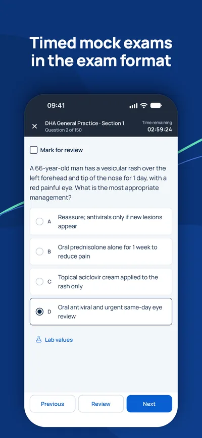
iPhone · iPad
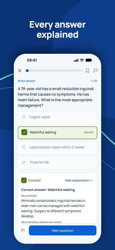
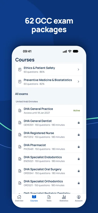
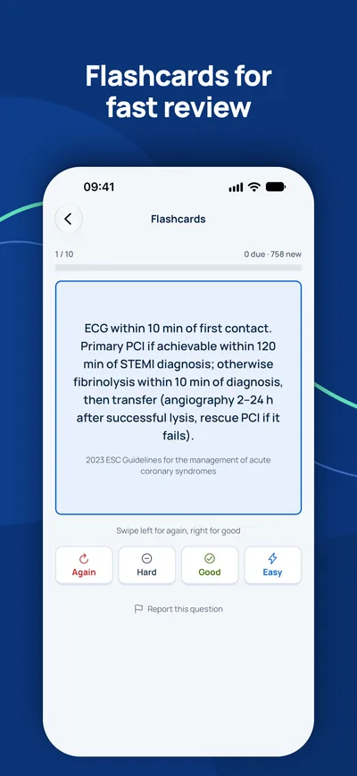
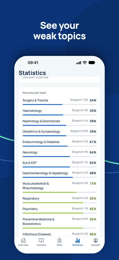
GCC Medical Exam Prep
Готовьтесь на iPhone к лицензионным экзаменам DHA, DOH, MOHAP, SCFHS и другим экзаменам стран Залива.


Экзамен — ваш единственный оставшийся шаг?
Сначала проверьте, нужен ли вам экзамен и на какое звание вы можете рассчитывать. Консультант ответит бесплатно.
Проверка соответствия- 1Бесплатная консультация по видео, телефону или WhatsApp
- 2Проверка соответствия официальным требованиям
- 3Ваш план с фиксированными ценами и понятными шагами